Anoxic Brain Injury
Findings:
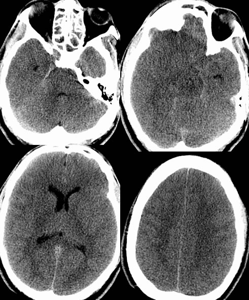
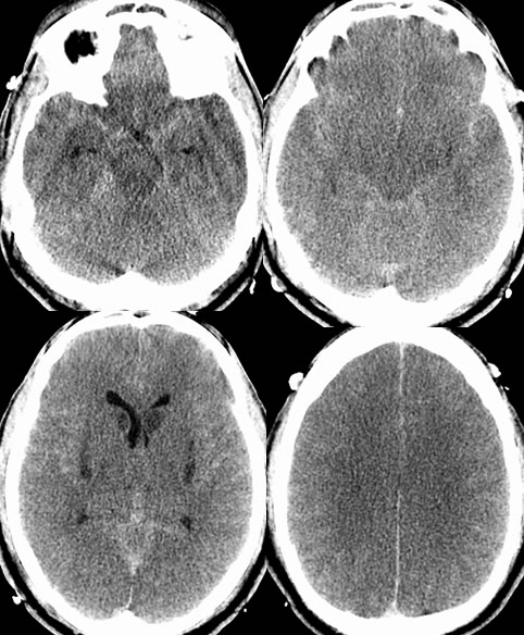
Multiple CT images demonstrate abnormal effacement of the basal cisterns and the third ventricle. Subsequent CT demonstrates development of focal areas of hypoattenuation in the bilateral insula and caudate nuclei. There is diffuse loss of grey white differentiation on the subsequent CT. The diffuse low attenuation brain parenchyma accentuates appearance of the leptomeningeal spaces, causing a pseudo-subarachnoid hemorrhage pattern.
Discussion:
While the initial CT findings are very symmetric and somewhat subtle for the uninitiated, the third ventricle and suprasellar cistern should be seen in all normal patients at any age. The lack of visualization of the third ventricle and/or suprasellar cistern usually indicates significant increased intracranial pressure. The subsequent CT confirms development of cerebral edema and bilateral infarcts.