Synovial Cyst
Findings:
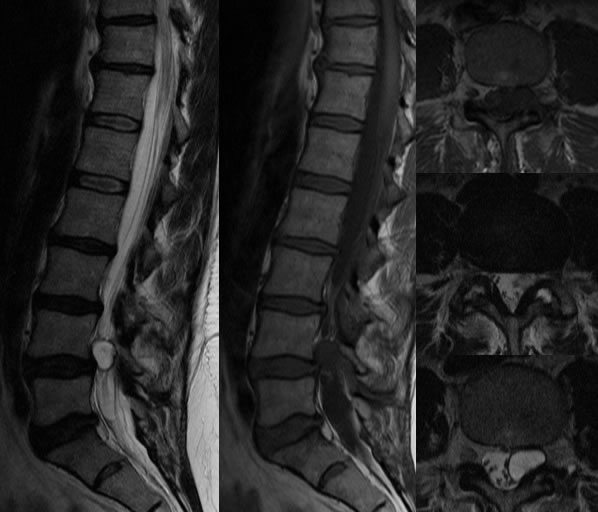
MR images of the lumbar spine demonstrates a well-defined cystic lesion with hypointense rim projecting from the left facet joint at L4-L5.The lesion causes significant compression left aspect of the thecal sac. There is also grade I anterolisthesis at L4-L5. A small left facet joint effusion is present.
Discussion:
nDifferential diagnosis
nDisc (anterior, less T2 hyper), ganglion cyst of LF (diff to distinguish), Nerve sheath tumor (enhancement), septic (soft tissue changes)
nImaging
nPosterolateral cystic lesion contig w facet joint, enhancing wall, hypointense rim, variable fluid signal usually follows CSF
nPath/Clinical
n90% lumbar, 70-80% at L4-5, connective tissue/synovium
nDegenerative, stress loading, facet instability, synovial proliferation
nRx
nConservative vs. surgical vs. percutaneous drainage and/or steroids
nBest lasting results w surgery if highly symptomatic
BACK TO
MAIN PAGE