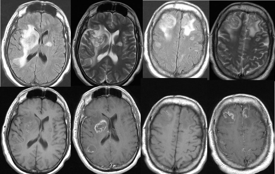
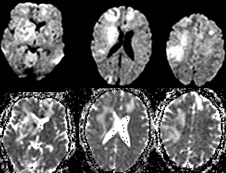
Toxoplasmosis
Findings:
Multiple MR images demonstrate multiple poorly defined enhancing lesions within the frontal lobes and basal ganglia.The lesions are associated with vasogenic edema with mild mass effect.The lesions also demonstrate some decreased patchy T2 signal suggestive of hemorrhagic changes. A mixed pattern of diffusion abnormality is associated with these lesions.
Discussion:
DDx: metastases, abscesses, multifocal glioma, lymphoma
-Most common cause of focal mass in HIV+