
Glioblastoma Multiforme
Findings:
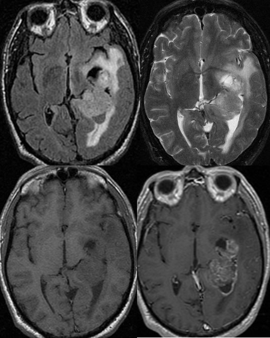
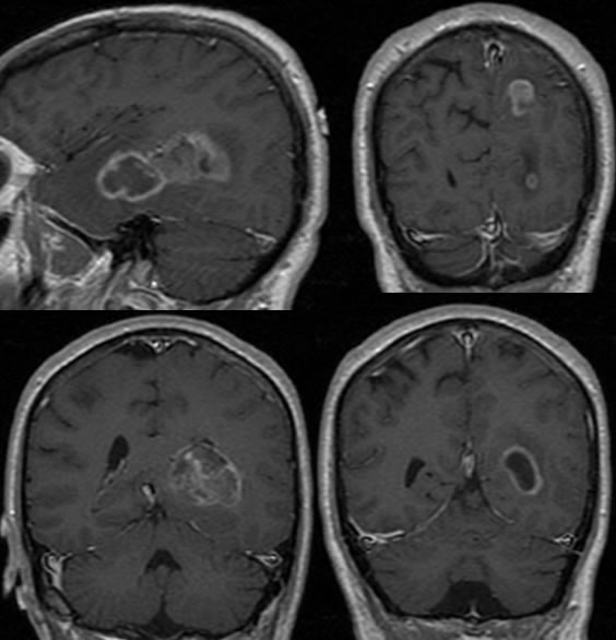
Multiple MR images demonstrate a poorly defined enhancing and nonenhancing mass involving the left temporal lobe with encroachment on the ambient cistern. There is also abnormal subependymal enhancement along the left lateral ventricle. Noncontiguous focus of abnormal enhancement is present in the left parietal lobe.
Discussion:
nmost common form of glioma, peak 45-55, M>F
ndeep frontal white matter most common, followed by temporal lobe and BG
nexpansile mass with central necrosis, ring enhancement, extensive edema, occasional calcification
nmicroscopic wide invasion despite well circ appearance
nddx: abscess rim typically hyper T1, hypo T2
nmay have flow voids
nGBM and lymphoma may spread across corpus callosum
nany T2 hyperintensity in CC means tumor spread, not edema
n8-12% 2ysr, c/w 38-50% for moderately anaplastic astro
BACK TO
MAIN PAGE