Septic facet arthropathy with epidural abscess
Findings:
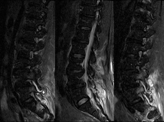
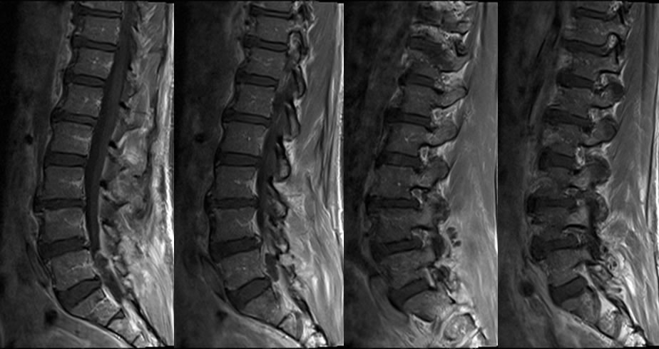
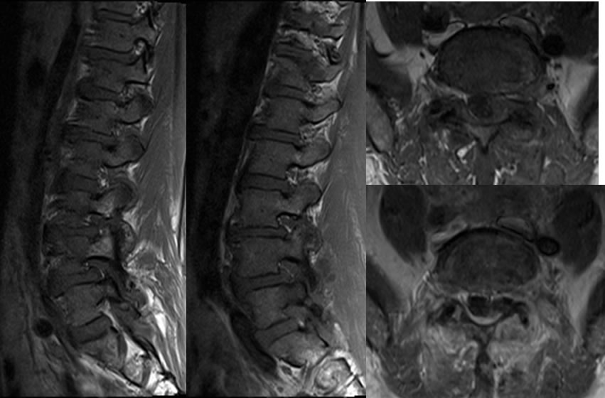
Sagittal T2 weighted images of the lumbar spine with fat saturation demonstrate extensive signal abnormalities within the paraspinal soft tissues at lower lumbar levels, with fluid collections within the facet joints, epidural space, and paravertebral soft tissues. There is abnormal marrow signal of the pedicles and facets at these levels. Postcontrast images confirm abnormal enhancement associated with this process with multiple fluid collections. An additional fluid collection with peripheral enhancement projects from the left L5 S1 facet, compatible with epidural abscess causing left thecal sac compression.
Discussion: