
Cerebral Amyloidoma
Findings:
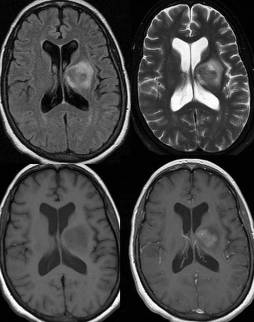
CT and MR images demonstrate a poorly defined somewhat flame shaped intraaxial mass within the left thalamus. The mass demonstrates marked T2 hypointensity with surrounding vasogenic edema and mass effect . The lesion is hyperdense on pre-contrast CT.
After radiation therapy and steroids, the mass effect has improved and the enhancing lesion is similar in size.
Discussion: