Schwannoma
Findings:
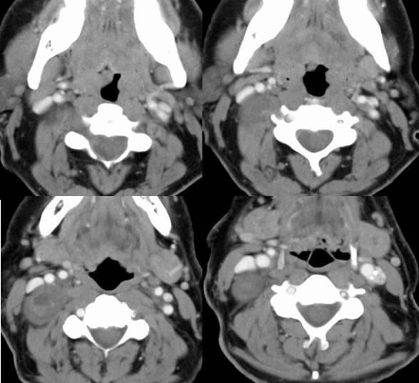
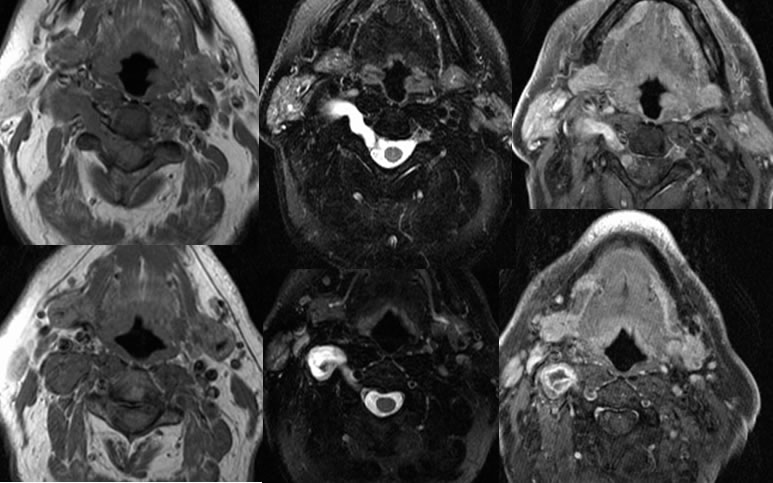
Multiple CT and MR images demonstrate a complex cystic and solid mass within the fat plane between sternocleidomastoid and scalene musculature. The mass demonstrates marked T2 hyperintensity with some zones of hypointensity and has an extent into the adjacent cervical neuroforamen. The mass demonstrates peripheral enhancement with a central zone of absent enhancement. A fluid-fluid level is seen in this region on CT.
Discussion:
nDifferential Diagnosis
nNeurofibroma (uniform), Meningioma (rare extradural), meningocele (nonenhancing)
nImaging
n75% intradural extramedullary, 15% dumbbell intra and extradural, 15% extradural
nVertebral erosion, foraminal widening, cystic changes, T2 target sign (hypointense center, swirling), intense enhancement
nPathology
nSingle nerve fascicle- sporadic most common (still mutations of NF2 gene in 60%)
nNF2- chr 22q mutation, schwannomatosis- peripheral wo NF2, Carney complex- melanotic schwannoma, chr 17, myxomas (cardiac), adrenal masses
BACK
TO MAIN PAGE