Meningioma
Findings:
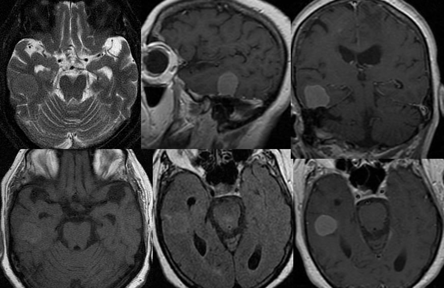
Multiple MR images demonstrate a homogeneously enhancing extra-axial mass projecting superiorly from the right petrous apex. There is no surrounding vasogenic edema. The mass is nearly isointense on FLAIR and T2/T1 weighted imaging.
Discussion:
n-Often hypo T2 due to fibrous content or calcification, strongly enhance, iso noncon T1. broad dural base, extraaxial, +/- dural tail.
n-common locations convexity, interhemispheric, tent. Classically iso to GM on all sequences.
-most common extraaxial neoplasm of adults, 15% of primary intracranial neoplasms, peak 50-60, F 2:1
-etiology unknown- ?trauma, radiation, virus, familial
-origin=arachnoid cap cell, possible assn with chr 22 deletion (9%- MISME)
-hormonally sensitive- pregnancy increases size
-malignant/aggressive more common in peds (imaging can't distinguish)
-dural tail (60%)-nonspecific, +/-cysts, +/- fat
-vascular supply: ECA 85%, ICA 63%
nGrading WHO 1993
meningioma I
atypical meningioma II
papillary, HPC, anaplastic II-III
BACK TO
MAIN PAGE