Glioblastoma Multiforme
Findings:
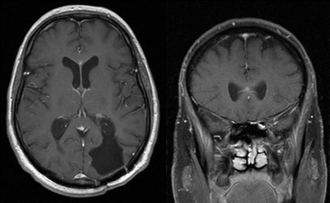
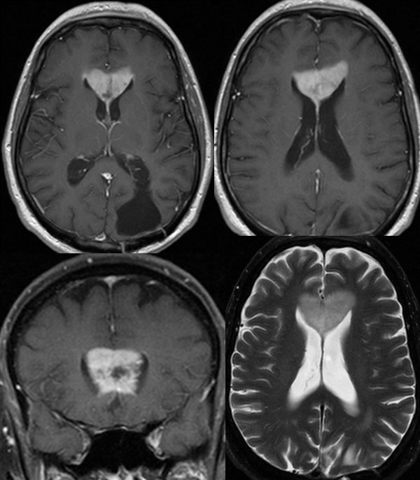
Multiple MR images demonstrate a lobulated mass near midline which demonstrates increased T1 and decreased T2 signal with surrounding FLAIR hyperintensity in both frontal lobes. Additional MR images confirm that the dominant mass arises within the corpus callosum with a contiguous poorly defined enhancing centrally necrotic mass involving the right frontal parasagittal cortex.
Discussion: