Osteomyelitis left temporal bone
Findings:
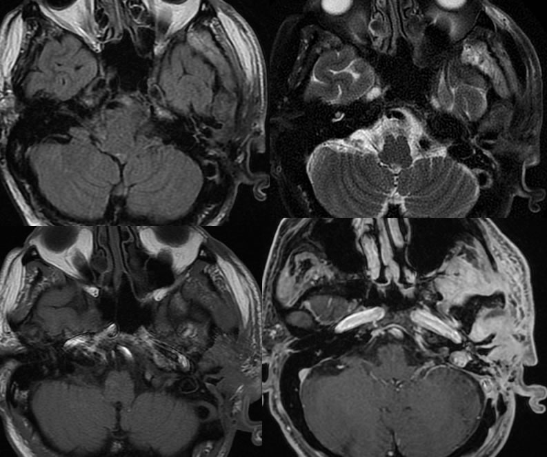
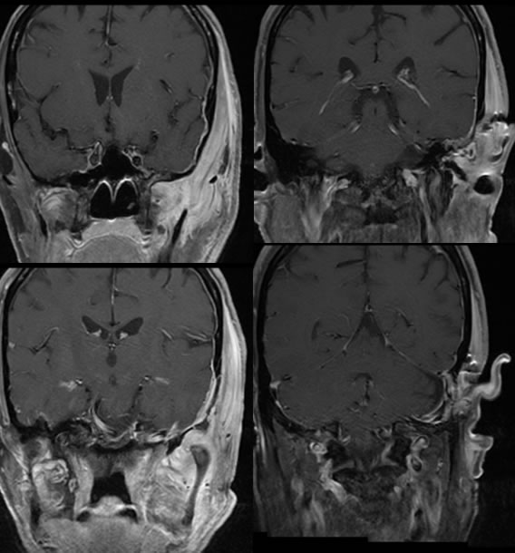
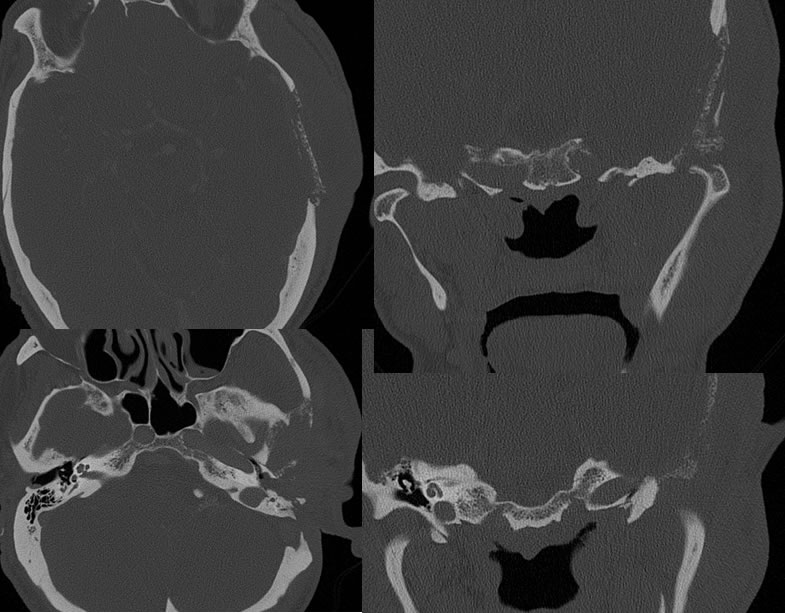
Multiple MR images demonstrate extensive infiltrative enhancement within the left masticator space and perimastoid region, extending into the condylar fossa. The process also surrounds the left mandible. CT images demonstrate extensive permeative destruction of the left squamous temporal bone and mastoid region including the condylar fossa. A destructive defect is present in the left mandibular condyle. Multiple irregular bony sequestra are present in this region.
Discussion: