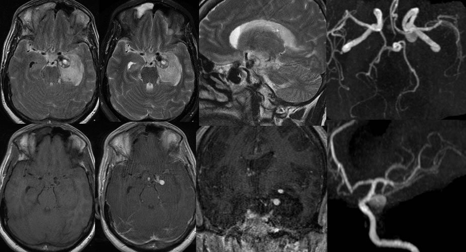
Left posterior communicating aneurysm with active rupture during scanning
Findings:
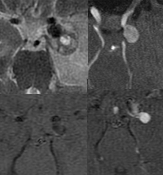
MR images without and with gadolinium demonstrate a large parenchymal hematoma in the left temporal lobe associated with subarachnoid hemorrhage. Extensive intraventricular hemorrhage is also present. The MRA reformatted images demonstrate a large aneurysm involving the communicating segment of the left internal carotid artery. On post gadolinium images, a globular focus of marked hyperintensity is noted adjacent to the aneurysm sac and within the superior aspect of the temporal lobe hematoma, compatible with active extravasation during the scan.
Differential Diagnosis:
Ruptured aneurysm, enhancement within aneurysm sac with sluggish flow, temporal lobe mass of other cause. With the presence of subarachnoid, parenchymal, and intraventricular hemorrhage as well as a definite aneurysm on CTA, etiologies other than aneurysm are not applicable. The distinction of aneurysm margins versus extraluminal extravasation can be made when the images are closely matched.
Discussion:
The communicating segment of the internal carotid arteries is the most common site of aneurysm along with the anterior communicating artery region. Extravasation during the scan is rarely seen and indicates emergent neurosurgical intervention unless other complications and clinical features preclude surgery. Further discussion of aneurysms is found on multiple other cases on this site.