Grade 3 Astrocytoma
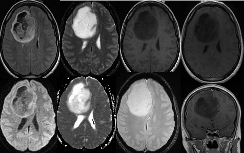
Findings:
Multiple MR images demonstrate a large heterogeneous lesion within the right frontal lobe. The mass demonstrates no definite abnormal enhancement or diffusion restriction. There is associated mass effect with frontal horn effacement and subfalcine herniation. The gradient echo sequence demonstrates no significant internal hemorrhagic change.
Differential Diagnosis:
Grade 3 astrocytoma, oligodendroglioma. GBM, abscess, and metastasis are not considered due to the lack of enhancement.
Discussion:
Discussion of glial neoplasms is found on many other cases on this site. Grade 2 astrocytomas are typically a homogenous, infiltrating, and mildly expansile lesion isolated to one lobe of the brain. As the lesions become higher grade, more heterogeneity, cystic portions, and enhancement are expected.