Ventral Cord Herniation
Findings:
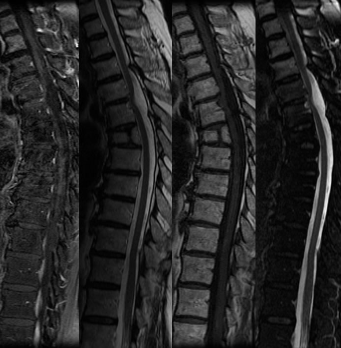
Sagittal MR images through the thoracic spine demonstrate abrupt ventral deviation of the spinal cord at the T5 level, which appears to invaginate into the dorsal margin of T5. A partial butterfly vertebra is seen at T6. The fat saturated post contrast image demonstrates no abnormal enhancement.
Differential Diagnosis:
Ventral cord herniation, subarachnoid adhesions, dorsal arachnoid cyst. Arachnoid cysts less typically cause an abrupt deviation but may coexist with VCH. Other epidural or intradural processes are not considered due to no enhancement and no abnormal signal within the CSF or epidural space.
Discussion:
Patients with VCH present with chronic unexplained progressive leg pain and myelopathy, with weakness, spasticity, Brown-Sequard syndrome, and sphincter dysfunction. The spinal cord focally herniates through a ventral dural defect which may be acquired or congenital, the exact etiology is not well established. These are most common at upper to mid thoracic levels and are characterized by an abrupt ventral deviation of the cord with no perceptible ventral CSF cleft. VCH is a rare but curable cause of myelopathy, treated with dural repair or enlargement of the defect to free the cord. An extradural sac may be present on occasion which has delayed filling on myelography with a filling defect representing the herniated cord. In the above case, the spinal cord herniates directly into the vertebral body which is rare but indicates a very long standing process.