Differential Diagnosis:
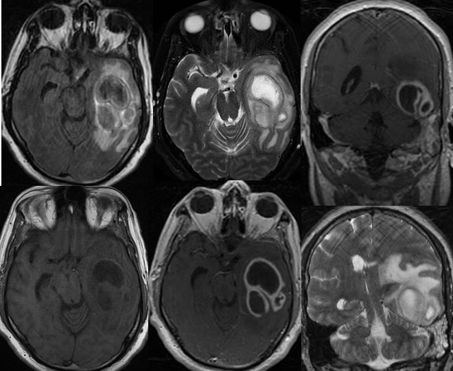
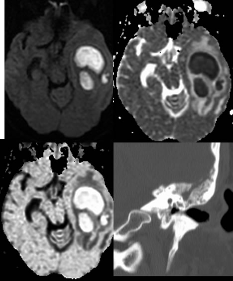
Abscess, high grade glial neoplasm, metastasis. Restricted diffusion and rim characteristics are key distinguishing features that indicate a diagnosis of abscess rather than the other possibilities. The presence of an obvious source in the left mastoid region also effectively eliminates the other possibilities.
Discussion:
Discussion of abscess is found in many other cases on this site. Importantly, 30% or more cases of abscess do not have an identifiable source. Headache and elevated ESR are fairly universal, but fever is found in only about 50%. Drainage is usually performed if greater than 2.5 cm. Any development of restricted diffusion in the previously drained cavity indicates recurrence.