Recurrent GBM, Corpus Callosum
Findings:
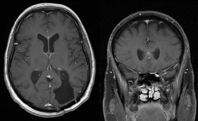
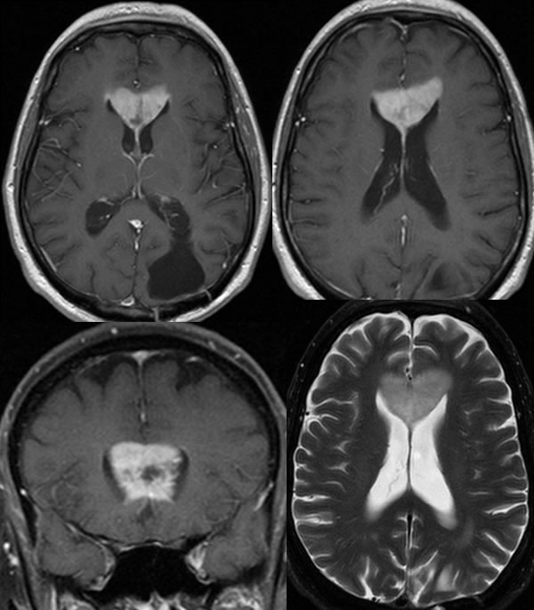
Axial and coronal T1 post contrast images demonstrate a surgical defect in the left parietooccipital region with overlying craniotomy. A tiny focus of enhancement is present at midline involving the anterior corpus callosum. Three months later, a large irregular enhancing mass has developed in the anterior corpus callosum compatible with recurrent tumor.
Discussion:
This case illustrates the importance of careful review of follow up studies in patients with GBM, where stable findings may be seen for several scans and a small subtle signal abnormality may develop that is easy to overlook. When a tiny new signal abnormality is overlooked, it is quite common to have explosive growth of the lesion within a few months that is obvious on the next follow up scan. Tiny areas of enhancement may be difficult to distinguish from pulsation artifacts, therefore careful coregistration to multiple planes is necessary. This punctate focus of enhancement is also somewhat difficult to note as an abnormality due to its precise midline location.