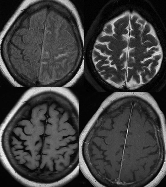
T-0
T- 2 weeks
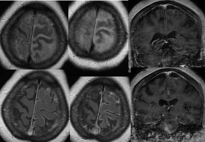
Meningoencephalitis
Findings:
Multiple MR images demonstrate abnormal FLAIR hyperintensity within left frontoparietal sulci that progresses over two weeks. On the post contrast images, there is peripheral leptomeningeal and cortical enhancement.
Discussion/Differential Diagnosis:
The differential diagnosis of FLAIR hyperintense CSF is broad, including subarachnoid hemorrhage, meningitis, leptomeningeal metastases, artifact, hyperoxygenation, or gadolinium in CSF.
Meningoencephalitis is usually caused by hematogenous seeding of the CSF from a more distant infection, is less commonly be caused by direct extension (sinuses, mastoids, orbits), and is rarely caused by penetrating injury. The organisms are most commonly bacterial such as Strep pneumo and N meningitides. Gram negatives and Listeria may also be seen in elderly. Other organisms include viral, TB, Cryptococcus, and Coccidioides. Complications include hydrocephalus, abscess, empyema, and infarcts. Infarcts may be caused by associated arteritis or venous thrombosis.
BACK TO
MAIN PAGE