
Glioblastoma Multiforme
Findings:
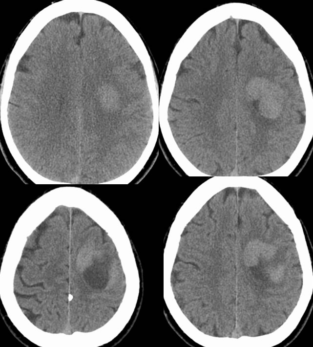
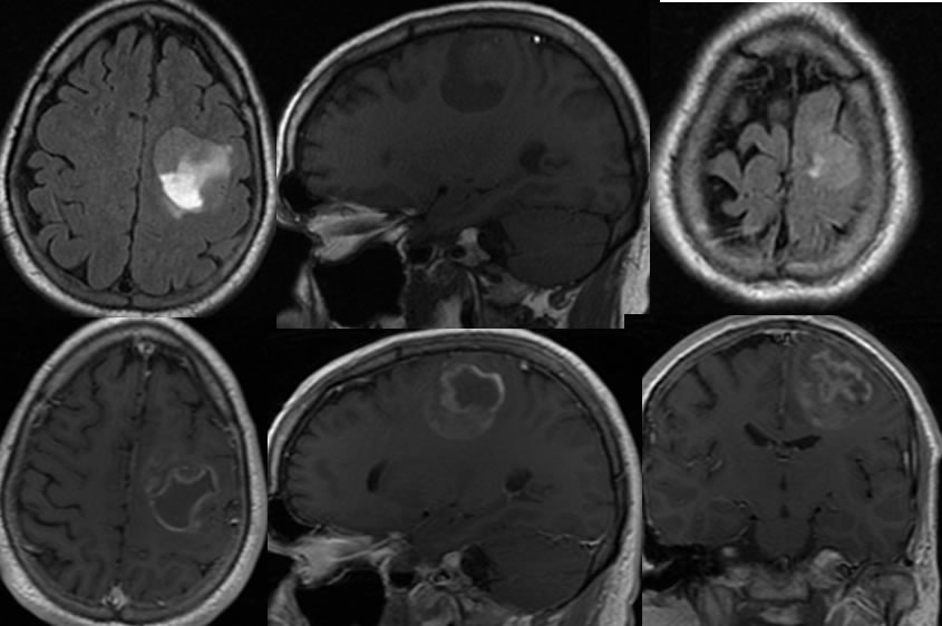
Axial noncontrast CT images demonstrate a heterogeneous lesion within the left frontal lobe that demonstrates mixed areas of homogeneous nodular hyperattenuation and cystic appearing zones of low attenuation. There is very little mass effect for the size of the lesion. The MR images demonstrate heterogeneously enhancing lesion in this region which causes only subtle distortion of the left lateral ventricle. There is significant swelling of the left superior frontal gyrus. Zones of FLAIR hyperintensity are present within the lesion and there is very little surrounding edema.
Discussion/Differential Diagnosis:
DDx: GBM, grade 3 astrocytoma, oligodendroglioma. The lack of significant mass effect and the overall signal characteristics would make abscess and metastasis unlikely. appearance would be very atypical for metastasis.
Please refer to other cases on this site for discussion of GBM. This case illustrates that GBM may be associated with less than expected mass effect for the size of the lesion since it may infiltrate with little reaction. It also illustrates the bizarre signal characteristics that may occur with GBM, with hyperdense areas on CT related to cellularity. The presence of gyral swelling suggests dedifferentiation of a previous lower grade astrocytoma.
BACK TO
MAIN PAGE


