Intraocular Melanoma
Findings:
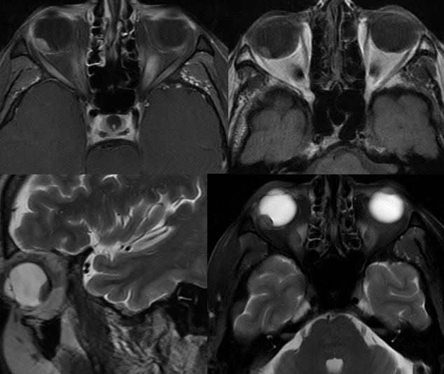
A enhancing circumscribed T2 hypointense, T1 hyperintense lesion is present in the inferolateralquadrant of the posterior segment of the right globe measuring approximately 10 x 9 x 5 mm (craniocaudal by greatest transverse dimensions).
Discussion/Differential Diagnosis:
The relative low T2 signal distinguishes from hemangioma and presence of enhancement differentiates this from retinal detachment. It is more focal and nodular than would be expected for an inflammatory lesion. Intraocular melanoma is the most common primary ocular malignancy in adults and is the second most common type of melanoma. Early detection is key to survival, and lesions located more peripherally from the optic disc present at a later stage. The lesions are initially detected on clinical eye examination and are first characterized by ultrasound. MR is helpful for detection of extent and metastases. Most arise from malanocytes in preexisting nevi, with genetic predisposition, sun exposure, and light colored iris representing additional risk factors. Worsening prognosis correlates with size and extent at detection. Epithelioid and/or amelanotic histology also portends a more aggressive course. Small indeterminate nevi may be followed with precise sonographic measurements, but larger lesions are treated with some combination of surgery and radiotherapy. Helpful MR imaging features for diagnosis include T1 hyperintensity, T2 hypointensity, and enhancement. Enhancement is the most consistent feature.
This case was in part prepared by Josh Hall, UC undergraduate.
BACK TO
MAIN PAGE