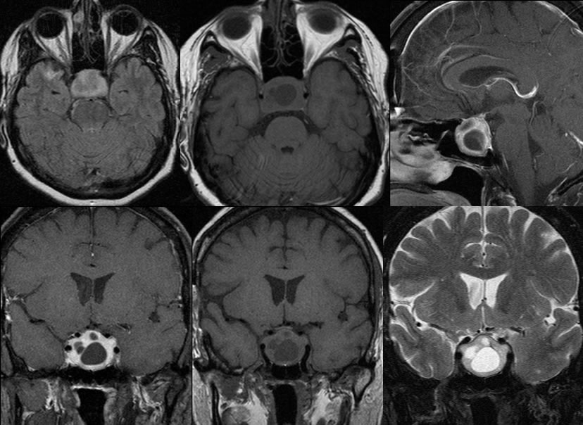
Pituitary Adenoma
Findings:
A large complex cystic and solid mass is present within the sella which measures 2.6 cm craniocaudal, 3.1 cm sagittal, and 2.4 cm AP dimension. The pituitary infundibulum is buckled and significantly deviated to the right. The suprasellar extent of tumor is inseparable from the optic chiasm and causes mild subtle mass effect and upward bowing of the optic chiasm. The mass extends into the medial margin of the left cavernous sinus and abuts the left cavernous internal carotid over a distance of approximately 180 degrees. There is also approximately 180 degrees abutment of the left supraclinoid ICA. The pituitary tumor also extends in the medial margin of the right cavernous sinus and abuts the right cavernous internal carotid over a distance of approximately 180 degrees. Discrete normal pituitary tissue is not visible. The largest cystic component in the sella measures up to 1.4 cm
Discussion/Differential Diagnosis:
DDx: Intrasellar craniopharyngioma (more common suprasellar and uncommon purely sellar, more cystic), metastasis (no known primary), meningioma (pit gland usually seen separate, solid lesion) and hyperplasia are solid. Not compatible with aneurysm.
Since up to 75% are hormonally active, pituitary adenomas often present with endocrine abnormality, or ">bitemporal due to chasm compression by suprasellar component. The imaging appearance isranging from complex cystic/solid to solid, variable size and signal but usually T1 isointense. Hemorrhage is uncommon and calcification is less common. Large masses may have a snowman configuration due a constriction at the diaphragm sellaeThey may be well defined with smooth sellar expansion, or more infiltrative with sellar and skull base destruction. Rare pituitary carcinoma may be indistiguishable from benign infiltrative adenomas. Enhancement is seen, but typically less than normal pituitary tissue. ICAs may be encased but not typically narrowed, unlike meningiomas involving the cavernous sinuses. Edema may be seen in the optic tracts with compressive lesions. They are 10-15% of intracranial tumors and most commonly prolactin secreting. They may be a part of the MEN 1 syndrome. They are treated surgically, with recurrence not uncommon. Metastases are very rare. Nonfunctional masses may be more aggressive with a higher recurrence rate.
This case was prepared with the assistance of Joshua Hall, UC undergraduate.
BACK TO
MAIN PAGE

