Osteoid Osteoma
Findings:
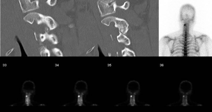
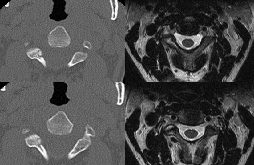
CT demonstrates a small lucent lesion with central nidus located in the right inferior articulating process of the C2 vertebra which measures approximately 5 x 4 x 4 mm in size. Intense uptake is present in this region on NM bone scan, with surrounding marrow edema signal on MR.
Discussion/Differential Diagnosis:
Often presenting in (3:1) male patients ages 10-35, osteoid osteomas (OO) are benign lesions that often present with nocturnal pain. This pain is most often treated by taking medication such as aspirin and surgical resection, and may also clinically present with minor swelling in the soft tissue surrounding the area. OO is an important cause of painful scoliosis in adolescents with concavity of curvature on the side of the lesion.
Up to 80% are found in long bones of limbs, about 20% in the phalanges, and 10% in posterior elements of the vertebrae (59% lumbar, 27% cervical, 12% thoracic, 2% sacral). On CT, a characteristic central ovoid lucency less than 2 cm will be seen with possible sclerosis/mineralization of the central nidus, associated with a fading gradient of sclerosis along the margins. It is important to note that the fading sclerosis is not part of the lesion but rather represents reactive changes.
Case prepared in part by Joshua Hall, UC undergraduate.
BACK TO
MAIN PAGE