Mucoepidermoid Carcinoma
Findings:
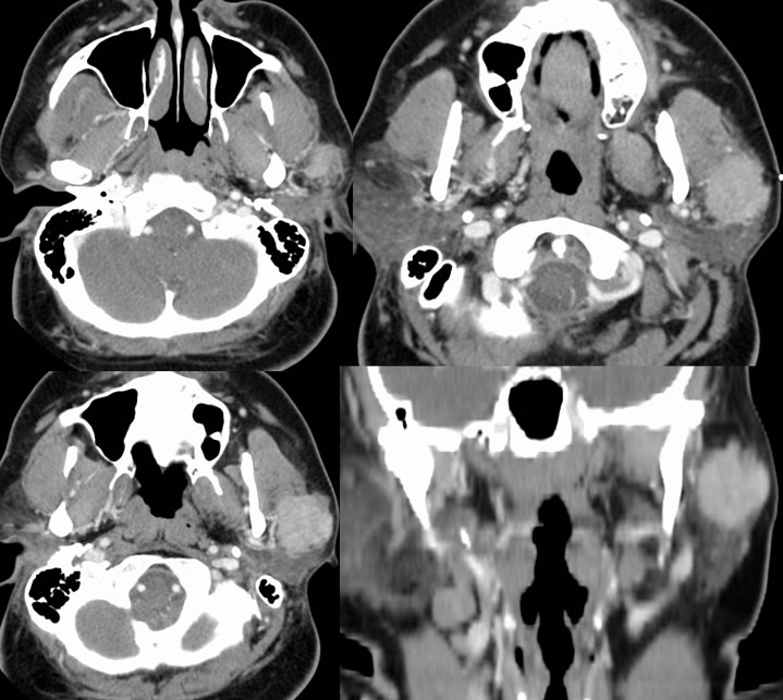
Contrast enhanced CT demonstrates a poorly defined irregular enhancing mass within the superficial lobe of the left parotid gland. The mass is inseparable from the adjacent masseter muscle and encroaches on the subcutaneous fat.
Discussion/Differential Diagnosis:
Differential diagnosis: Pleomorphic adenoma, mucoepidermoid carcinoma, adenoid cystic carcinoma, lymphoma, metastasis.
While about 80% of parotid masses are benign such as pleomorphic adenoma (AKA benign mixed tumor- BMT) or Warthin's tumor, certain features such as irregular margins, local invasion, and adenopathy should raise the possibility of a more aggressive lesion. With aggressive lesions, the search should continue through the expected retromandibular course of the auriculotemporal nerve and stylomastoid notch for soft tissue infiltration that may indicate perineural tumor spread of malignancy and worsened prognosis.
Mucoepidermoid carcinoma (MEC) are less than 10% of salivary gland tumors but comprise 30% of salivary gland malignancies with up to 50% occurring in the parotid. MEC may also arise in minor salivary gland tissue, are the most common pediatric salivary gland malignancy, and the second most common submandibular gland malignancy behind adenoid cystic. The histologic grade correlates with prognosis, with low grade lesions well circumscribed and indistinguishable from BMT by imaging. The low grade lesions are usually solid but may contain cystic areas and calcification is rare. High grade MEC is generally more homogenous with infiltrating margins and hypercellular characteristics on MR (low-intermediate T2 signal). The histologic grade also dictates the extent of surgery, with wide local excision for low grade and radical resection and neck dissection/XRT for high grade lesions. Recurrence is not uncommon for higher grade lesions. Histologically, there is a prominent mucinous component and goblet cells intermixed with epithelioid squamous like features. Typical findings of high mitotic rate, necrosis, and nuclear pleomorphism are seen with high grade lesions.
Case contributed by Josh Hall, UC undergraduate
BACK TO
MAIN PAGE