Pleomorphic Adenoma, minor salivary gland
Findings:
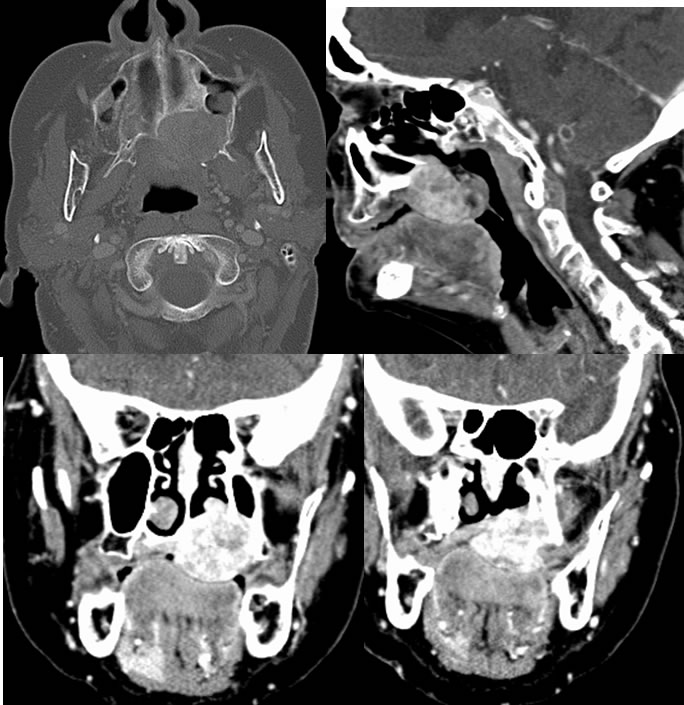
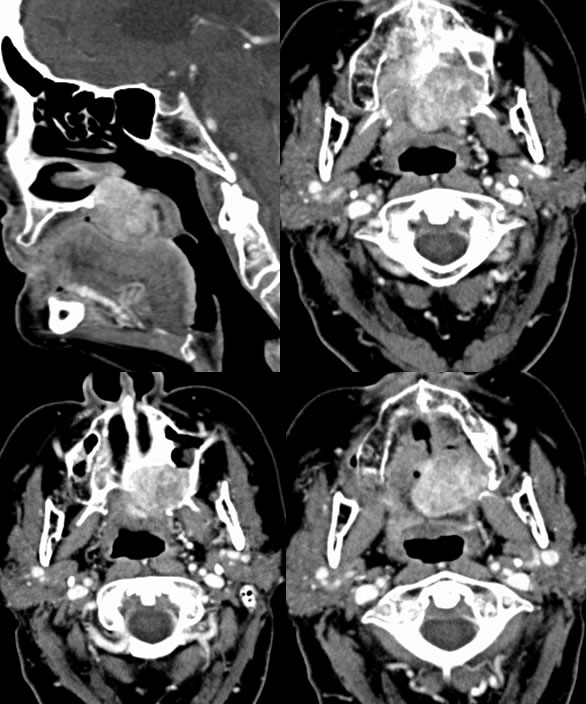
A large heterogeneous strongly enhancing mass involves the left hard palate which is associated with destruction of the medial pterygoid process and posterior medial margin of the left maxillary sinus, destruction of the left hard palate. There is also subtle bone remodeling in this region with a thin sclerotic border in some regions indicating a relatively long-standing process. This mass has an approximate transverse dimension of 3.5 x 2.3 cm, with a craniocaudal dimension of 2.5 cm. On the coronal reconstructed images, the mass extends slightly to the right of midline with destruction of the medial aspect of the right hard palate as well as a small focus of inferior posterior nasal septum destruction. It extends exophytically into the superior aspect of the oral cavity and also has a small lobulated extent into the floor of the nasal cavity through the hard palate defect.
Discussion/Differential Diagnosis:
Differential Diagnosis:Benign mixed tumor, squamous cell carcinoma, lymphoma, schwannoma
Benign mixed tumors (BMT) are the most common salivary gland tumors with almost all occurring in the head and neck region. Up to 85% are found in the parotid gland, with less than 10% found in the submandibular gland and oral/pharyngeal mucosal spaces. Less than 1% are found in the sublingual glands. When small, BMT may be indistinguishable from more aggressive histologies including squamous cell carcinoma, lymphoma, and minor salivary gland malignancy. The imaging appearance is nonspecific and heterogeneous, ranging from solid enhancing to complex cystic and solid, and occasionally partially calcified. Benign tumors are typically well circumscribed but may scallop bone if large.
The appearance of this mass is nonspecific, but the bone remodeling indicates a long standing process, which should raise suspicion for a low grade slow growing lesion such as pleomorphic adenoma, also known as benign mixed tumor. Of the possibilities above, the enhancement is atypical for schwannoma and this is less aggressive than would be expected for squamous cell carcinoma or lymphoma. A more homogenous lesion might be expected for lymphoma.
BMT arise from myoepithelial tissue associated with salivary gland tissue anywhere along the upper aerodigestive tract and have a diverse cellular morphology. Depending on their location, BMT of mucosal spaces may present with painless obstructive symptoms and/or dysphagia. BMT are treated surgically with an excellent prognosis, but can reach massive size if ignored. Malignant transformation of BMT may occur, up to 10% when it involves minor salivary gland tissue. If left untreated, up to 25% will eventually undergo malignant transformation.
This case was prepared with the assistance of Joshua Hall, UC undergraduate
BACK TO
MAIN PAGE