Hemorrhagic pineal cyst
Findings:
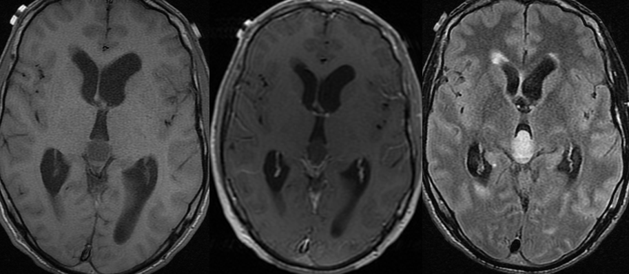
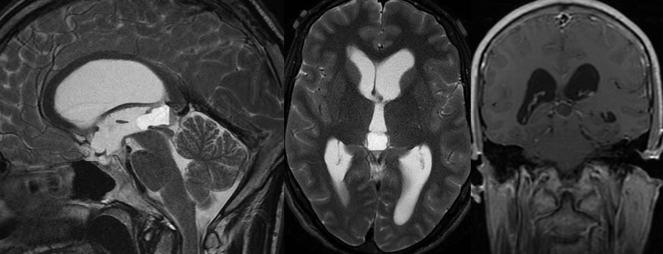
There is an ovoid mass centered in the pineal measuring 2.2 cm AP by 1.3 cm transversely by 1 cm. There is a fluid/fluid level. It is predominantly T1 hypointense, T2 hyperintense. The layering fluid has susceptibility artifact on gradient. This does not enhance. There is mass effect on the proximal aspect of the cerebral aqueduct. There is moderate dilatation of the lateral and third ventricles with mild periventricular T2/FLAIR hyperintense signal, right greater than left.
Discussion/Differential Diagnosis:
Differential Diagnosis: Hemorrhagic pineal cyst, pineocytoma (nodular component expected), arachnoid cyst, germinoma (solid expected). Epidermoid expected to be more irregular with DWI restriction, also rare in this location.
The vast majority of cystic pineal glands are benign incidental findings of no clinical significance. When these lesions are less than 1 cm but reported in the impression, this may lead to long term probably unnecessary follow up MR imaging. They are typically less than 1 cm in size but may be larger and more heterogeneous causing compression of the tectum and cerebral aqueduct with hydrocephalus, as in this case. Nodular and more heterogeneous components may be present, making them sometimesindistiguishable frompineocytoma by imaging.Embyologically, they arise from failure of obliteration of the cavum pineal, with debris and/or hemorrhagic products sometimes accumulatingand causing a more complex appearance. They may enlarge with hormonal stimulation, and are most common in females (3:1). Acute hemorrhage with rapid expansion (pineal apoplexy) is rare but may be fatal.
This case was prepared with the assistance of Joshua Hall, UC undergraduate
BACK TO
MAIN PAGE