
Pineal Germinoma
Findings:
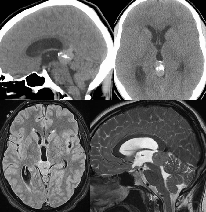
On CT, a hyperdense mass with coarse predominantly central calcifications in the pineal region measures approximately 2.3 x 1.2 cm in greatest axial dimensions and extends approximately 2.7 cm in craniocaudal dimension. Sagittal reformatted images suggest superior displacement of the internal cerebral vein. There is effacement of the cerebral aqueduct, and resultant moderate enlargement of the lateral and third ventricles. On MR images, the mass demonstrates homogeneous enhancement, slight T1 precontrast hypointensity, and intermediate T2 signal. The intermediate T2 signal suggests dense cellularity. The anterior margin of the mass is infiltrative and inseparable from the medial thalami and third ventricle. The mass is inseparable from the vein of Galen.
Discussion/Differential Diagnosis:
Differential Diagnosis: Germinoma, Pineocytoma, Meningioma, Glioma, Metastasis, all generally indistinguishable when solid and homogenous.
Germ cell tumors such as germinoma are overall uncommon, less than 2% of primary brain tumors. Other types of intracranial germ cell tumors include benign and malignant teratomas, choriocarcinoma, endodermal sinus tumor, embryonal carcinoma, and mixed histologies. Germinoma is the most common intracranial germ cell tumor and the most common pineal tumor, demonstrating histology identical to testicular seminoma. The pineal germinomas are typically seen with a male predominance (10:1), age 10 to 20, and are far more common in Asia than North America. Germinomas may also be found in the suprasellar region or uncommonly in the thalami or basal ganglia, but are nearly always near midline. Patients present with obstructive hydrocephalus and Parinaud syndrome (upward gaze palsy and altered convergence) if in the pineal region. Suprasellar germinomas may present with endocrine symptoms including diabetes insipidus and precocious puberty as well as bitemporal visual field defects related to chiasmal compression.
Germinomas are hyperdense on CT due to cellularity and tend to engulf the pineal calcification as opposed to pineocytomas which expand the pineal calcification. They enhance homogeneously. Hemorrhage and cystic changes are rare. Obstructive hydrocephalus may be seen due to compression of the aqueduct. The tumors are usually nearly isointense to gray matter on precontrast sequences. Subependymal and/or subarachnoid spread may be seen and should be sought on MR. Fortunately, Germinomas are highly radiosensitive and potentially curable with resection and radiation therapy, with greater than 90% fysr. Somatic malignant transformation (sarcoma, adenocarcinoma) is rare but portends a very poor prognosis. The prognosis also worsens for mixed histologies.
This case was prepared with the assistance of Joshua Hall, UC undergraduate
BACK TO
MAIN PAGE