Dermoid
Findings:
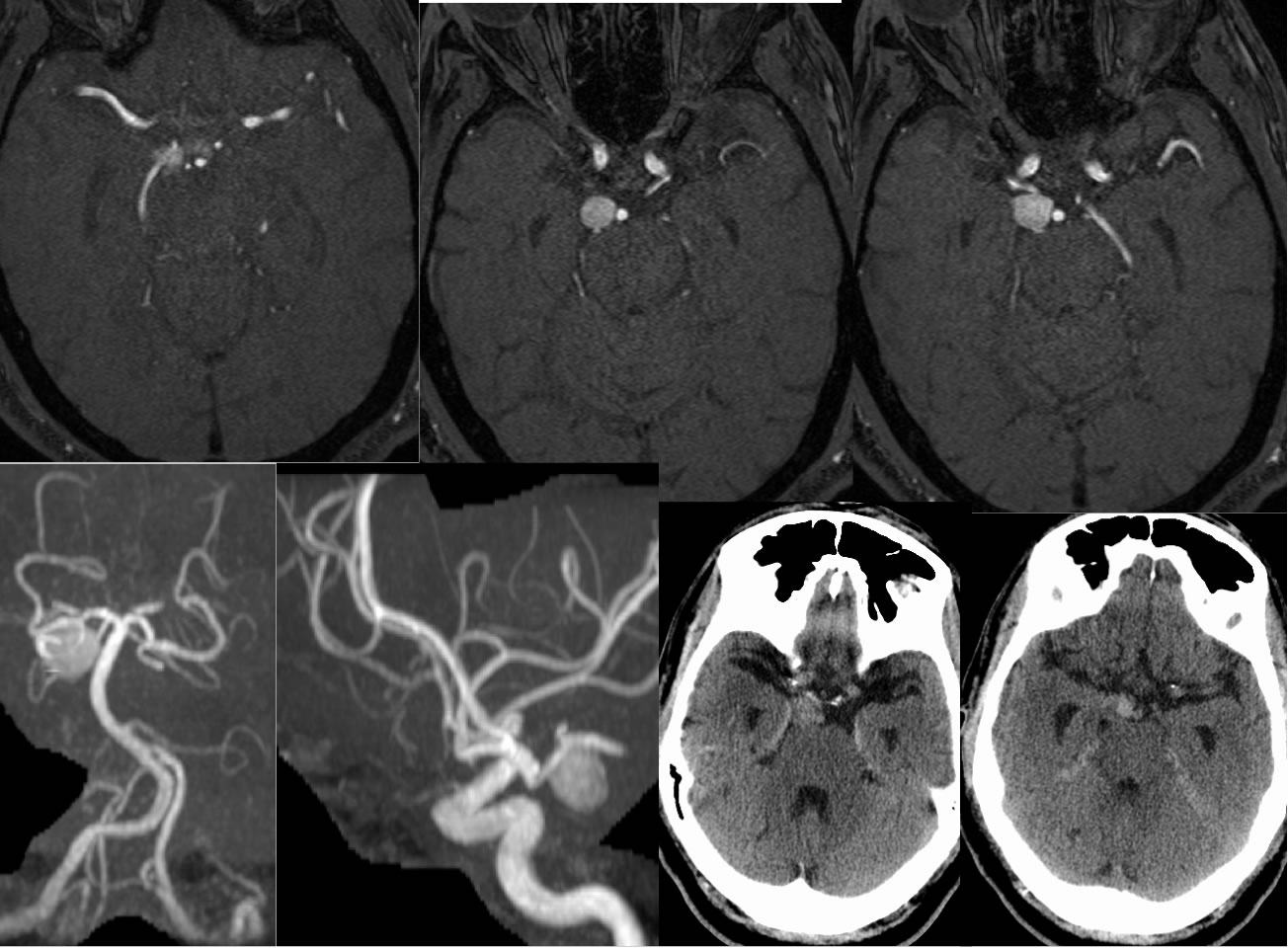
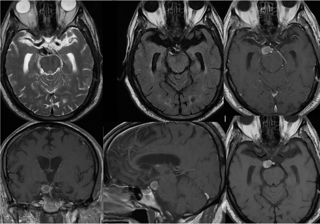
MRA source images demonstrate a round hyperintense lesion along the undersurface but not contiguous with the right posterior cerebral artery. The lesion is slightly less hyperintense than vascular flow on MRA. The lesion is faintly hyperdense on CT. It also demonstrates decreased T2 and increased T1 signal without definite abnormal enhancement. A small lobule of the lesion also extends along the anterior aspect of the basilar artery.
Differential Diagnosis/Discussion:
Partially thrombosed aneurysm is the top differential to exclude due to proximity to the PCA and round morphology, but the lack of pulsation artifact through the lesion and atypical signal characteristics for aneurysm indicate the more likely possibility of an extraaxial mass with complex proteinaceous signal (hyper T1, hypo T2) such as a dermoid. Other type of suprasellar cystic lesion could be considered, but not craniopharyngioma or other tumor due to location and lack of enhancement.
BACK TO
MAIN PAGE