CADASIL
Findings:
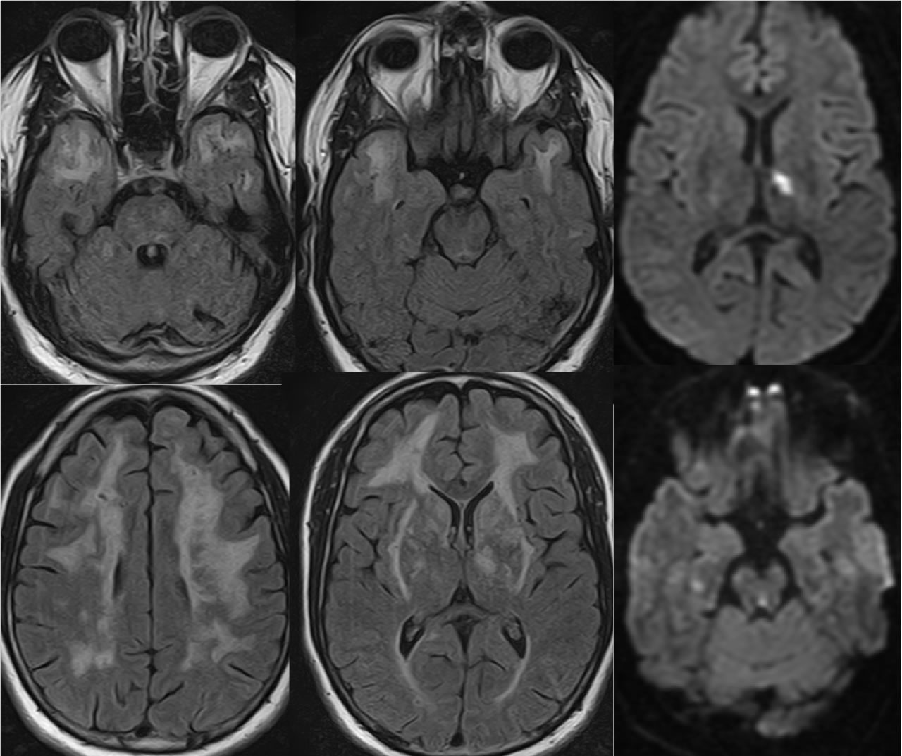
Multiple axial FLAIR images demonstrate extensive confluent white matter signal abnormalities in both cerebral hemispheres, which also extend into the anterior temporal white matter. These signal abnormalities also extensively involve the basal ganglia, caudate, and external capsule regions. A small remote infarct involves the left cerebellum. The diffusion weighted images demonstrate recent lacunar infarcts in the left thalamus, right hippocampus, and dorsal midbrain.
Differential Diagnosis:
While confluent white matter disease in general has a broad differential diagnosis that is narrowed by age of the patient and clinical features, the presence of patchy bilateral anterior temporal subcortical white matter signal changes as well as age likely less than 60 raises the possibility of CADASIL.
BACK TO
MAIN PAGE