Cerebellar AVM with feeding aneurysm of PICA
Findings:
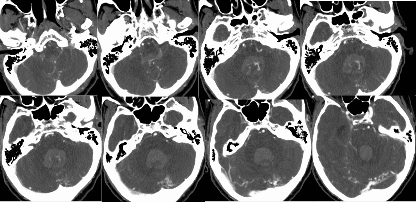
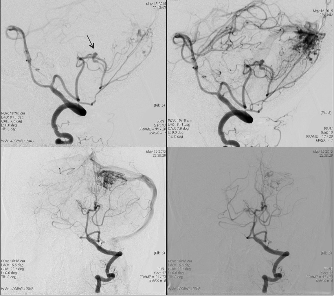
Axial CTA source images demonstrate intraventricular and subarachnoid hemorrhage in the posterior fossa. A cluster of abnormal vessels is present near the falx cerebelli. Left PICA is enlarged along the fourth ventricular margin. Conventional arteriogram demonstrates an irregular aneurysm of PICA which feeds the AV shunting lesion in the posterior fossa.
Differential Diagnosis/Discussion:
While the differential diagnosis for isolated posterior fossa hemorrhage is broad, the abnormal tortuous vascularity seen on CTA indicates an underlying shunting lesion as a cause. A feeding vessel aneurysm is one of many possible causes of hemorrhage in the presence of an AVM and are often difficult to detect without a conventional arteriogram.
BACK TO
MAIN PAGE