Hereditary Hemorrhagic Telangiectasias
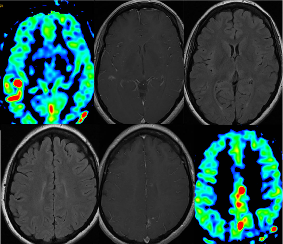
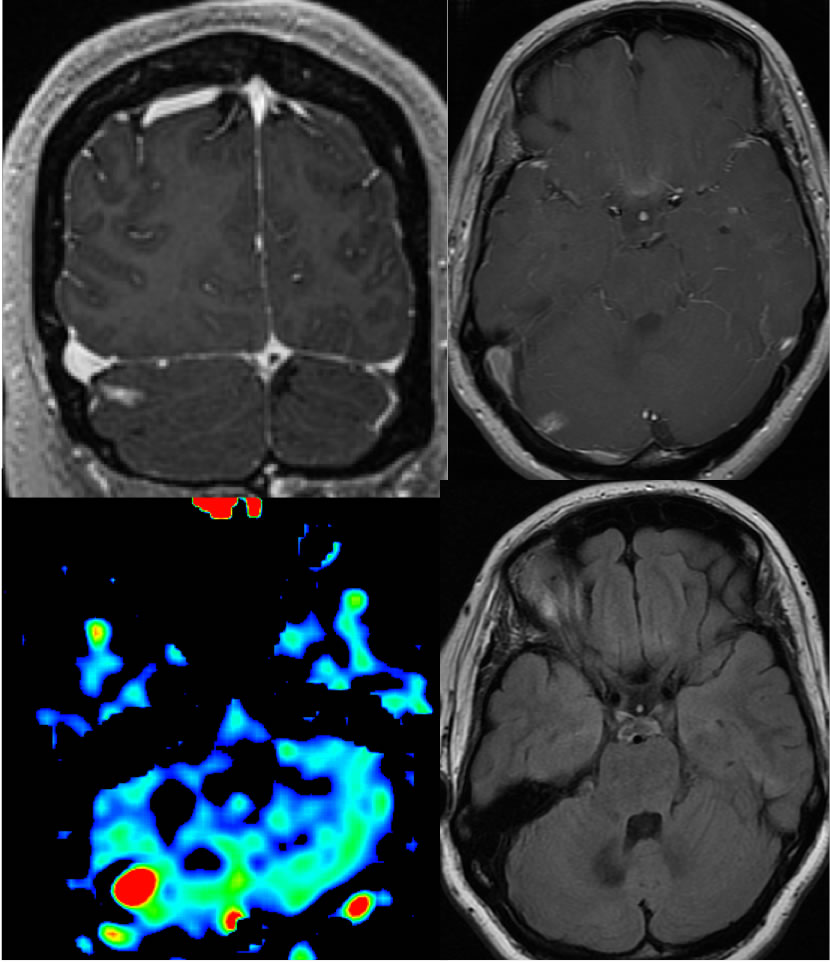
Findings:
Scattered irregular linear and bandlike foci of abnormal enhancement are present within the right lateral cerebellum, left frontoparietal parasagittal region, and right parietal lobe, which do not demonstrate corresponding FLAIR hyperintensity, but do demonstrate hyperperfusion on the ASL sequence.
Differential Diagnosis/Discussion:
The lack of nodularity, mass effect, edema, and absence of signal abnormality on precontrast sequences indicates a nonneoplastic cause. The presence of patchy enhancement in the absence of signal abnormality on other sequences is fairly typical for telangiectasias, and the presence of multiple lesions should raise the possibility of a congenital syndrome. Multiple subacute infarcts with fogging effect can occasionally appear similar, but would not be associated with hyperperfusion.
BACK TO
MAIN PAGE