Central Skull Base Osteomyelitis
Findings:
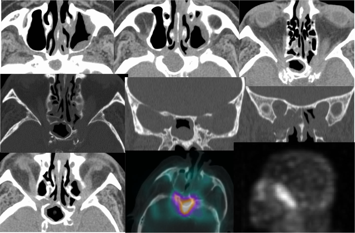
Moderate mucosal thickening is present in the sphenoid sinus, associated with surrounding sclerosis and irregular thickening of the sinus walls. Several bone defects are also present due to bone destruction. Abnormal soft tissue opacity is seen within the bilateral pterygomaxillary fisures, retromaxillary fat pads, and bilateral orbital apices left greater than right. Gallium scan shows intense increased uptake in this region.
Differential Diagnosis/Discussion:
While inflammatory mucosal thickening is very commonly seen within the paranasal sinuses, it is important to search for features that indicate a more aggressive infectious process. These features include bone destruction and adjacent soft tissue infiltration. The lack of a focal nodular mass would argue against malignancy. The pterygomaxillary fat planes are an important region to inspect for the presence of inflammatory or neoplastic infiltration.
BACK TO
MAIN PAGE