Progressive Multifocal Leukoencephalopathy
Findings:
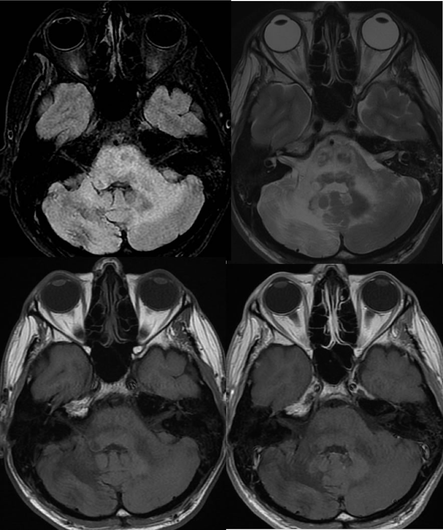
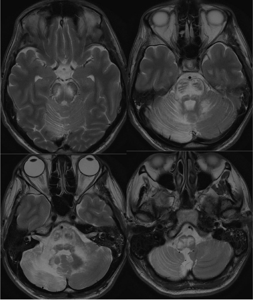
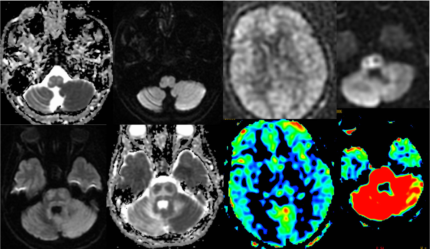
Multiple MR images demonstrate widespread confluent T2 hyperintensities within the cerebellum and brainstem, which notably spare the corticospinal tracts and are not associated with abnormal enhancement or mass effect. Slightly reduced ADC values are seen in some regions, but overall T2 shine through phenomenon is seen on the DWI. ASL perfusion demonstrates dramatic diffuse hyperperfusion of the affected areas.
Differential Diagnosis/Discussion:
PML with or without IRIS, gliosis from remote insults, other cerebellitis, ADEM. Infiltrating neoplasm unlikely without mass effect or enhancement. The presence of hyperperfusion is a helpful feature to distinguish PML from PML-IRIS, favoring the former (Khoury, et al Brain 2013 Nov;136(11):3441-3450.).
BACK TO
MAIN PAGE