Subdural Empyemas with Meningoencephalitis
Findings:
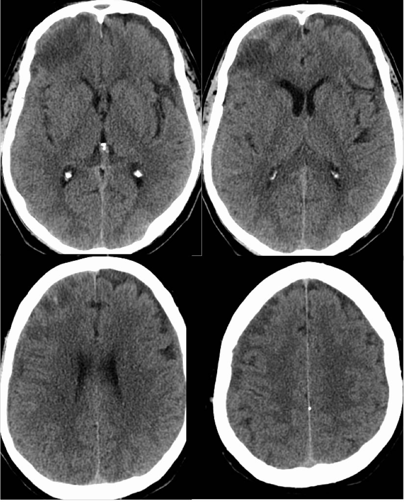
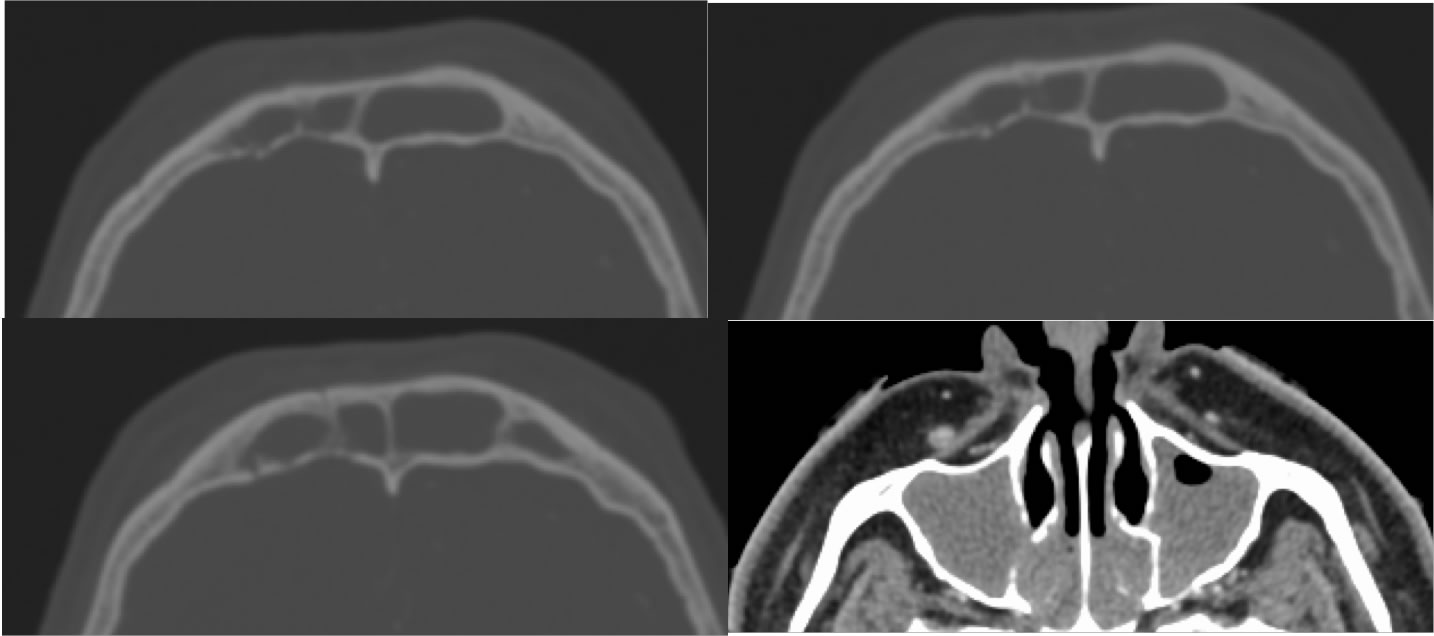
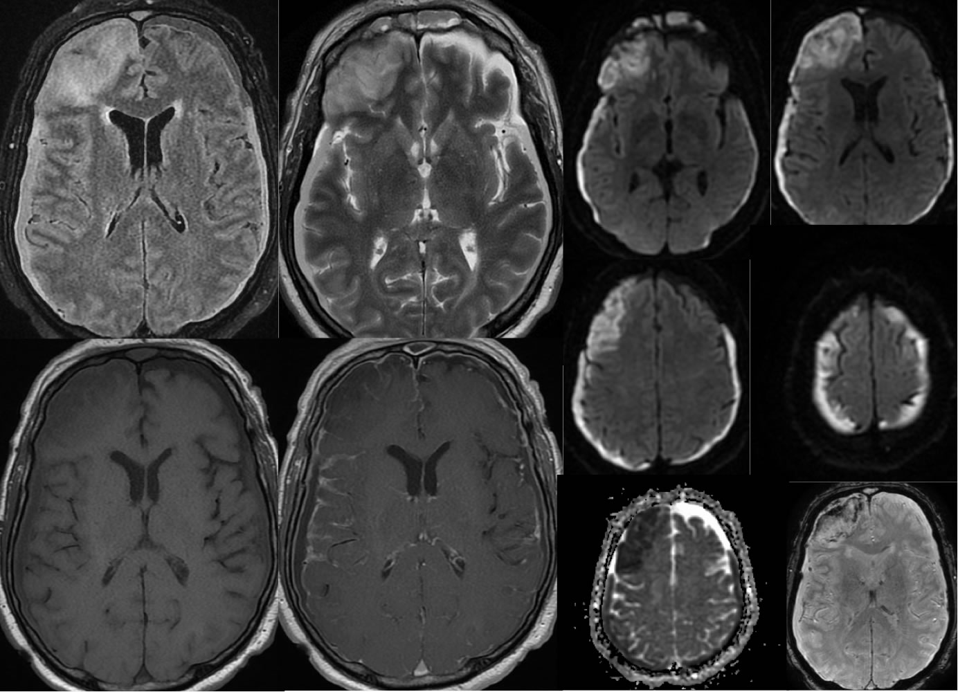
Noncontrast CT images demonstrate asymmetric patchy low attenuation within the right frontal lobe which also involve cortex. Complex bilateral subdural fluid collections are present with mixed density, causing subtle symmetric mass effect. Selected bone window images demonstrate marked opacification of the paranasal sinuses with demineralization of the frontal sinus walls. MR imaging demonstrates restricted diffusion within the bilateral subdural fluid collections, associated with leptomeningeal enhancement. There is also restricted diffusion within the right frontal lobe and spotty hemorrhagic changes. Abnormal marrow signal and enhancement of the frontal bones is completely included.
Differential Diagnosis/Discussion:
The noncontrast CT appearance was initially felt to represent aging contusion in the right frontal lobe and mixed density bilateral subdural hematomas, due to a stated history of trauma, however the presence of extensive sinus disease and erosive changes of the frontal sinuses does raise the alternative possibility of infection. The MRI confirms the intracranial infectious process, with the extensive restricted diffusion within the complex collections that is different in morphology than would be expected for the susceptibility due to hemorrhage. Encephalitis and underlying right frontal lobe infarction would be impossible to distinguish precisely.
BACK TO
MAIN PAGE