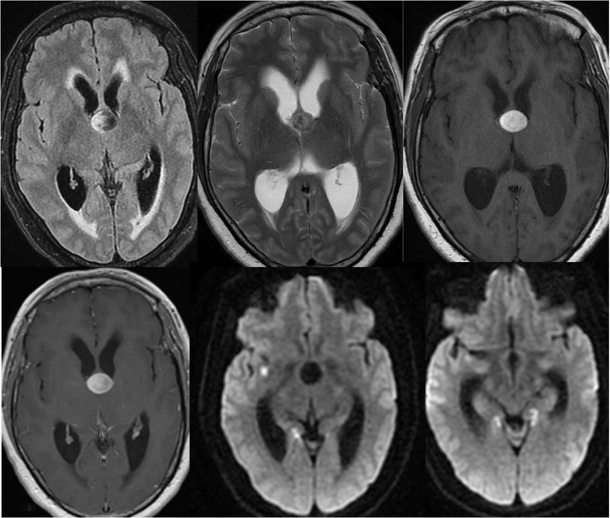
Colloid Cyst with bilateral hippocampus and right subinsular infarcts
Findings:
Multiple MR images demonstrate dilated ventricular system due to obstructive hydrocephalus at the level of the foramina of Monro. There is evidence of transependymal CSF flow with hazy periventricular T2/FLAIR hyperintensity. The causative lesion is a rounded mass at the left foramen of Monro which demonstrates mixed but predominately hypointense T2 and hyperintense T1 signal. No abnormal enhancement is present. Superimposed zones of diffusion restriction are present within the right subinsular region and bilateral hippocampi.
Differential Diagnosis/Discussion:
The differential diagnosis for obstructive lesions at the foramen of Monro include colloid cyst, central neurocytoma, subependymoma, giant cell astrocytoma (look for TS) , and other lesions including metastasis. The lack of enhancement indicates a non-neoplastic process, although subependymomas also may be nonenhancing. However, the well-defined nature, hyperintense T1 signal, and hyperintense T2 signal is fairly characteristic for colloid cyst.
BACK TO
MAIN PAGE