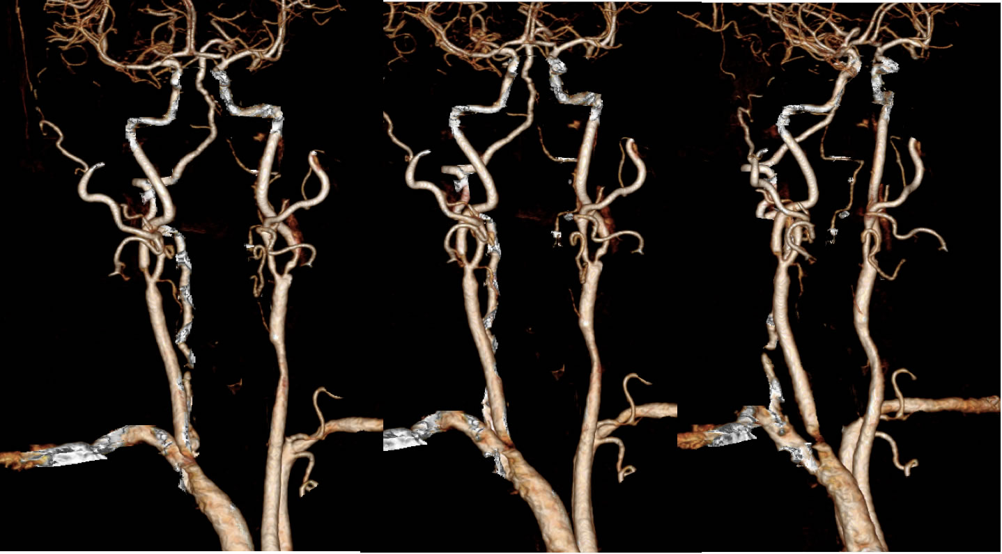
Severe Bilateral Internal Carotid Stenosis, L Vertebral Artery Occlusion, High Grade Stenosis R Vertebral Artery
Findings:
VRT CTA reformats show multifocal irregular stenoses of arterial structures. These include bilateral high-grade internal carotid origin stenoses, high grade stenosis of the proximal dominant right vertebral artery, and multifocal occlusion/high grade stenosis of the left vertebral artery. Irregular mild to moderate basilar artery stenosis is also seen. There is also a proximately 40-50% stenosis of the mid portion left common carotid.
Differential Diagnosis/Discussion:
Multifocal arterial stenosis are by far most commonly caused by atherosclerotic disease. Multifocal smooth vascular narrowings could also be caused by dissections or arteritis. Occasionally, tumor encasement may cause a smooth isolated vascular narrowing. Please note that VRT images cannot be relied upon for precise diagnosis due to underestimation of stenosis and inability to assess regions of heavily calcified disease. In this case, the stenoses are more irregular than would be expected for multifocal dissection or arteritis.
BACK TO
MAIN PAGE
