Odontogenic Abscess
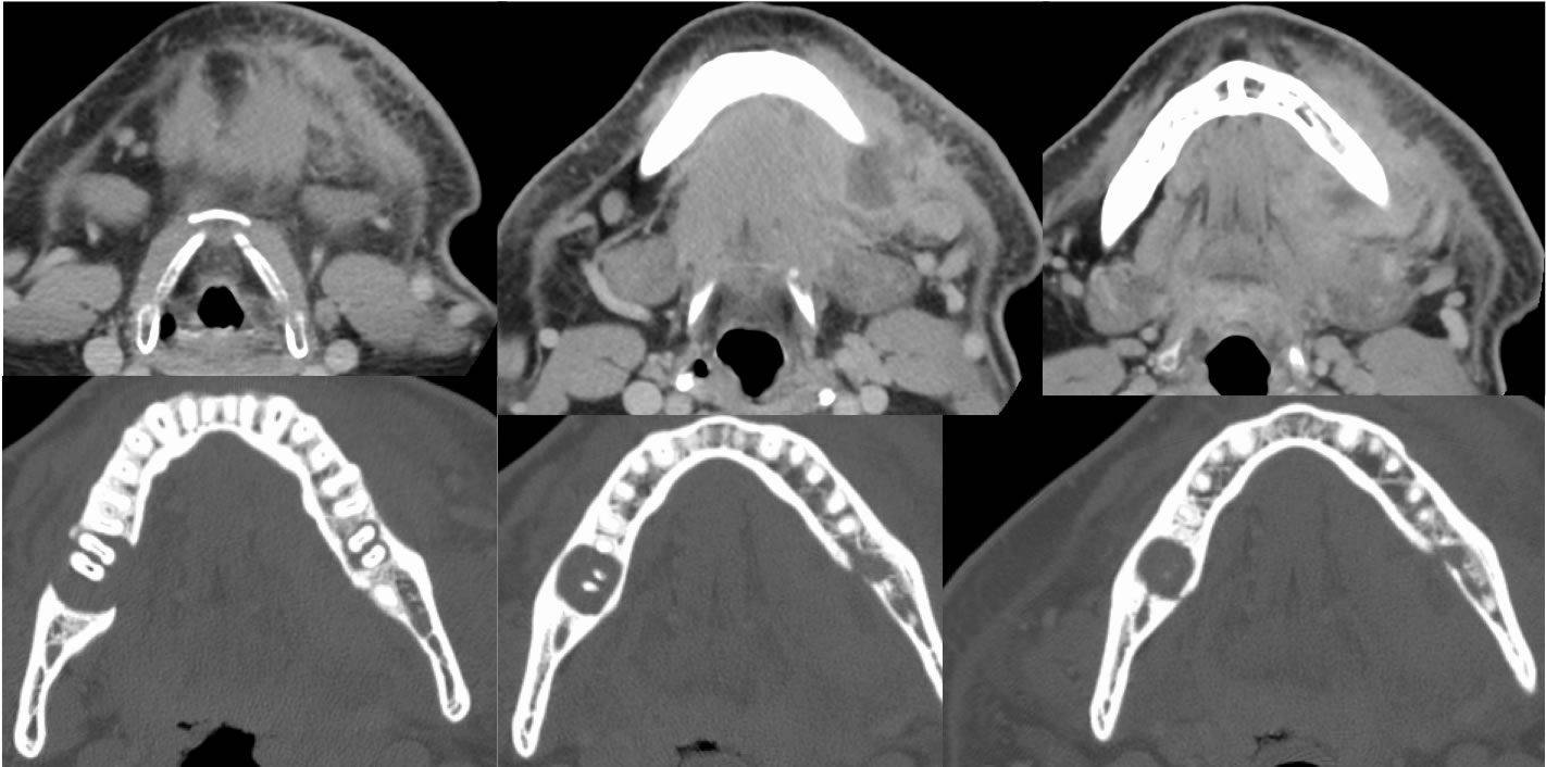
Findings:
Contrast enhanced CT images of the face including bone algorithm reconstructions show a poorly defined fluid collection surrounding the left mandibular body, associated with hazy surrounding soft tissue induration, thickening of the platysma muscle, and reactive adenopathy. The bone windows demonstrate a destructive defect along the medial margin of the left mandibular body associated with a periapical cyst of tooth number 19. A larger well-defined periapical cyst is associated with tooth number 31, but there is no active soft tissue inflammatory process currently visible in this region.
Differential Diagnosis/Discussion:
This is not usually a diagnostic dilemma since the patient will come into the ER with a toothache and facial swelling, or be sent from the dentist with the same. The associated abscess is easy to detect as a fluid collection with rim enhancement and surrounding inflammatory change. The offending tooth needs to be extracted, and often these patients have other zones of dental disease, therefore it is imperative to localize the culprit by associated periapical cyst and mandible destruction that is in the region of the soft tissue inflammatory process.
BACK TO
MAIN PAGE