Craniopharyngioma
Findings:
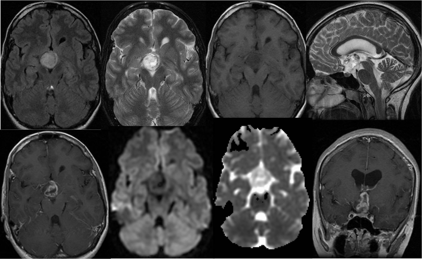
Multiple MR images demonstrate a complex cystic and solid suprasellar mass with irregular enhancement. The mass appears separate from the pituitary tissue. The mass is inseparable from the optic chiasm and floor of the third ventricle. The mass is well-defined without surrounding edema.
Differential Diagnosis/Discussion:
The differential diagnosis for suprasellar masses is broad, but pituitary origin lesions can generally be excluded if a normal pituitary gland is located. Ectopic pituitary tumor would be rare. For a complex cystic and solid supersellar mass in this region, primary differential diagnostic considerations include craniopharyngioma and hypothalamic/chiasmatic glioma. The absence of surrounding edema and infiltration of the hypothalamus indicates that the former is more likely. A metastatic lesion could appear similar if there is a known primary cancer. A germinoma is more likely a solid lesion, and enhancement would not be expected with a hamartoma.
BACK TO
MAIN PAGE