Perineural Tumor Spread, Squamous Cell Carcinoma
Findings:
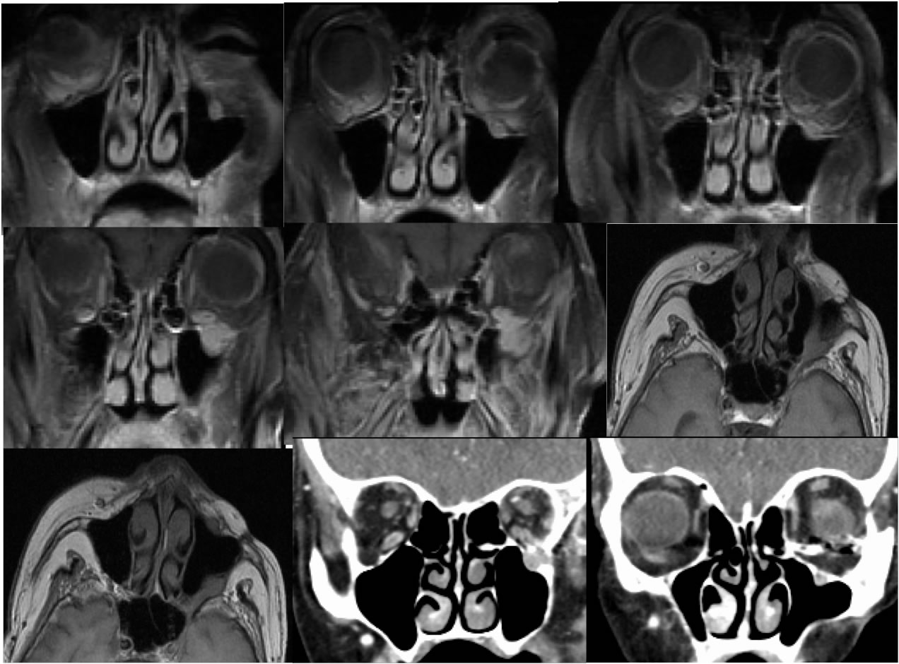
Coronal fat saturated T1 post contrast, axial T-1, and coronal contrast enhanced facial CT images demonstrate postoperative scarring in the left premaxillary region related to previous tumor resection. Cordlike enlargement of the left infraorbital nerve is present with a poorly defined enhancing mass along the posterior superior left maxillary sinus infiltrating the pterygomaxillary fissure. Loss of the pterygomaxillary fat plane is seen on the axial T-1 images. A tiny mass is also seen along the anterior left maxillary sinus wall on the T1 axial. Destruction of the maxillary sinus wall is less well visualized without bone reconstructed CT images.
Differential Diagnosis/Discussion:
Occasionally, infiltrative soft tissue at a postoperative site may be difficult to distinguish between recurrent tumor and postoperative scarring on imaging. However, an important task of the neuroradiologist is to detect the presence of perineural tumor spread which manifests as abnormal enlargement of a nerve supplying the primary tumor site, and infiltration of the deep facial structures including the pterygomaxillary fissure would not be expected for postoperative scarring and perineural tumor spread should be considered, in addition to the presence of a destructive mass that indicates malignancy.
BACK TO
MAIN PAGE