Tuberous Sclerosis
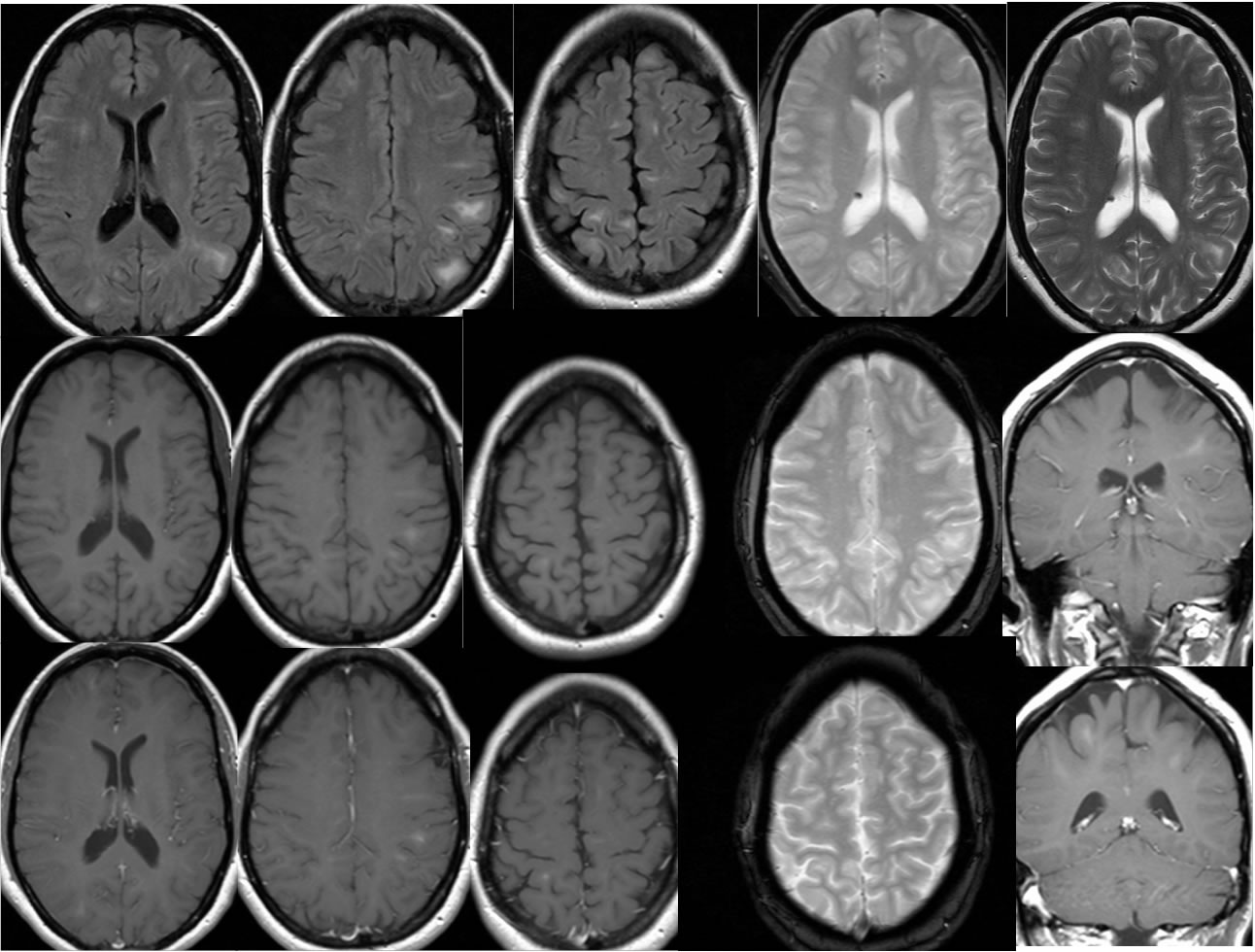
Findings:
Multiple MR images demonstrate scattered ovoid bilateral subcortical FLAIR hyperintensities, many of which are associated with T1 hyperintensity that may reflect calcification. No definite abnormal enhancement is seen and there is no hemorrhagic staining. A single hypointense subependymal nodule is present within the right lateral ventricle. Definite masses are not seen at the foramen of Monro.
Differential Diagnosis/Discussion:
The presence of calcified subcortical lesions and ependymal nodules should raise the possibility of Tuberous Sclerosis. Nodules near foramen of Monro need not be present for the diagnosis. Dystrophic calcifications related to other remote insults such as congenital infection would be less likely to be associated with patchy FLAIR hyperintensities.
BACK TO
MAIN PAGE