Metastatic Lung Cancer
Findings:
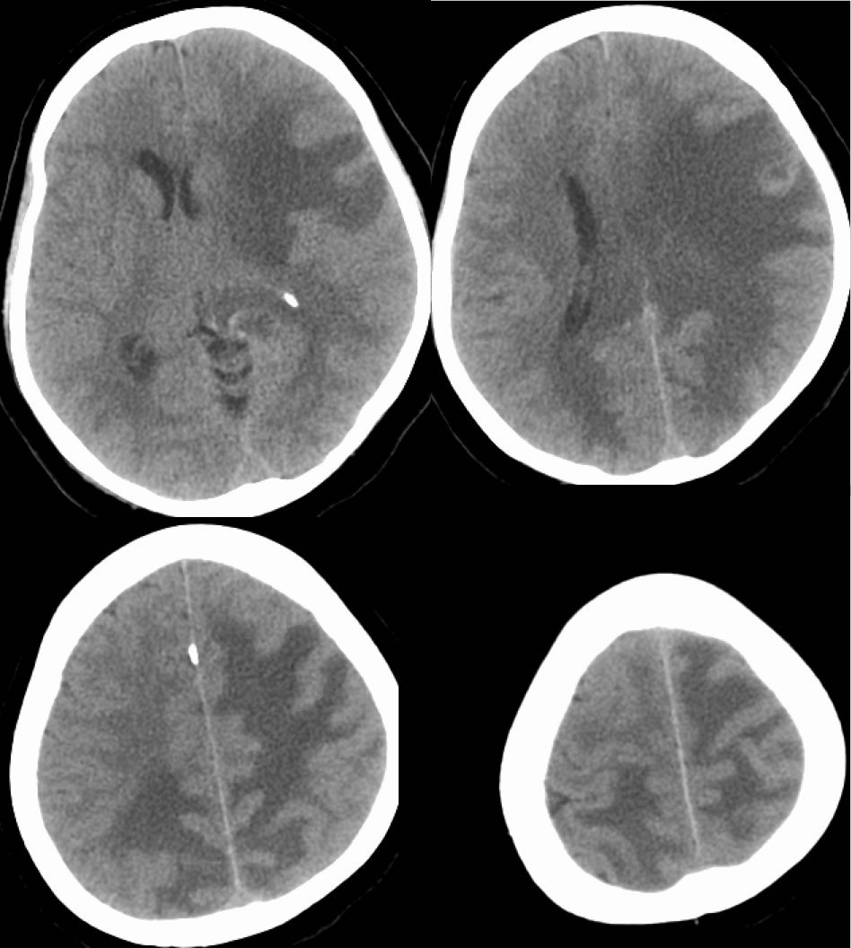
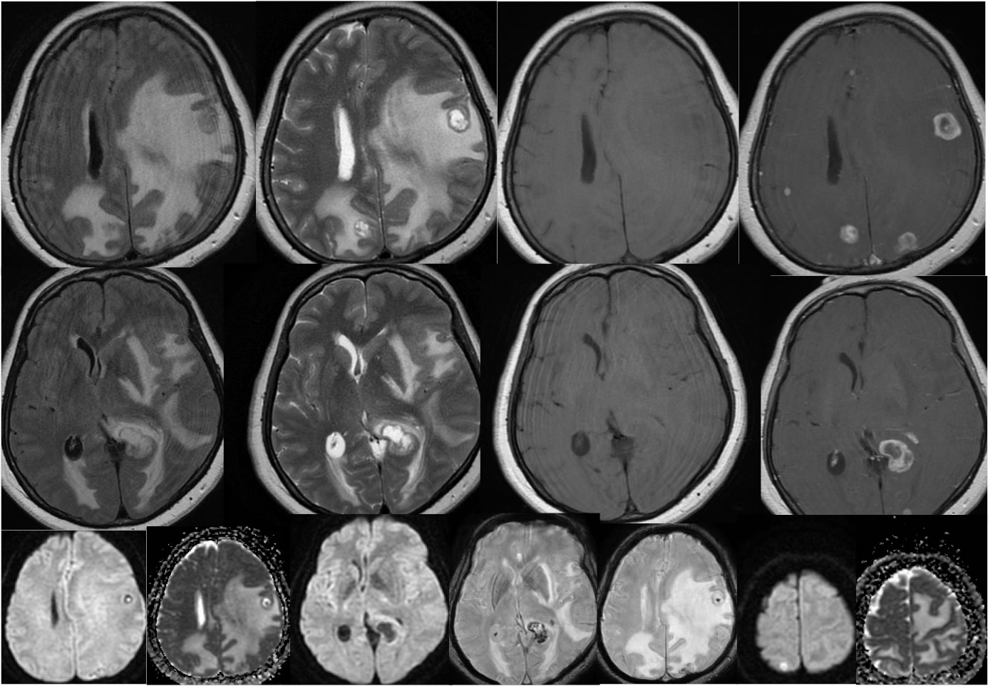
Axial CT images demonstrate extensive zones of vasogenic edema throughout the cerebral hemispheres left greater than right, associated with significant mass effect and left to right midline shift. One zone of nodularity is visible within the L frontal edema on CT. The MR images reveal multiple irregular ring enhancing lesions within the edema, some of which demonstrate hemorrhagic staining, but no central restricted diffusion.
Differential Diagnosis:
The differential diagnosis of multiple ring enhancing intraaxial lesions is broad, but by far most commonly related to neoplastic disease or infection. The absence of diffusion restriction in this case indicates that infection is less likely. The degree of irregular nodularity, hemorrhagic changes, and edema indicate the high likelihood of metastatic neoplasm. While multifocal primary glial neoplasm (GBM) and lymphoma can also cause multiple lesions, this degree of edema and mass effect would be unusual. The presence of these multiple lesions without obvious signs of infection should trigger a search for primary neoplasm, in this case chest CT demonstrated lung cancer.
BACK TO
MAIN PAGE