Anoxic Brain Injury due to Electric Shock
Findings:
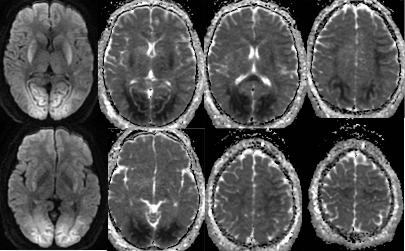
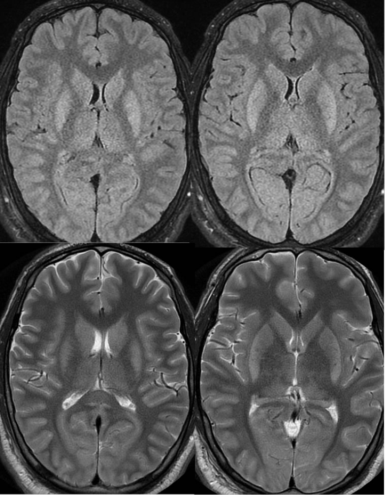
Extensive zones of restricted diffusion symmetrically involve the occipital lobes, basal ganglia, corpus callosum, and watershed zones of the bilateral cerebral hemispheres. While fairly subtle and extremely symmetric, associated hyperintensities are seen in these regions on the T2 and FLAIR images.
Discussion:
The symmetric appearance of the scan might cause the uninitiated to think this is normal or has some unusual artifact. Clinically, the patient has had poor neurological recovery after electrocution that caused ventricular fibrillation. CPR and defibrillation was required, but time to ROSC is not known. The imaging patterns of anoxic injury may be variable, but are often symmetric reflecting the diffuse insult. It is uncertain whether the decreased autoregulation of the posterior circulation contributed to this appearance.
BACK TO
MAIN PAGE