T 0
T 6 Months
Neuromyelitis Optica
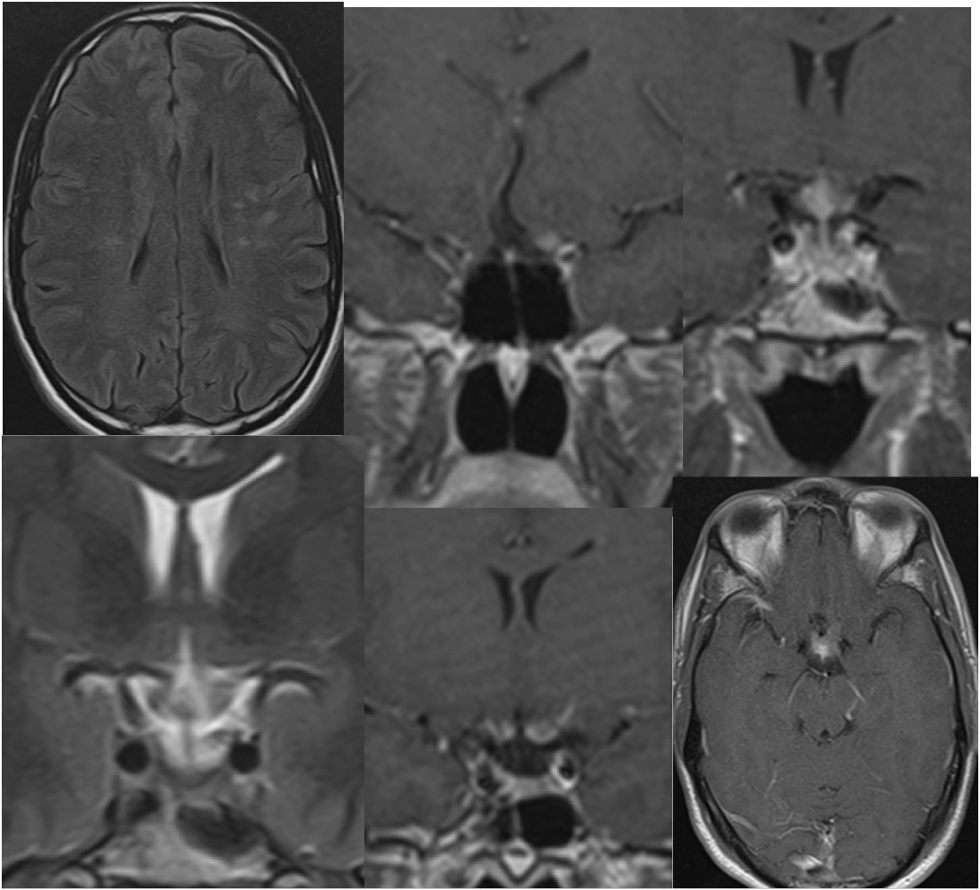
Findings:
The initial MRI images demonstrate scattered ovoid deep bilateral frontal white matter signal abnormalities, with expansion and abnormal patchy enhancement of the optic chiasm and proximal optic nerves.
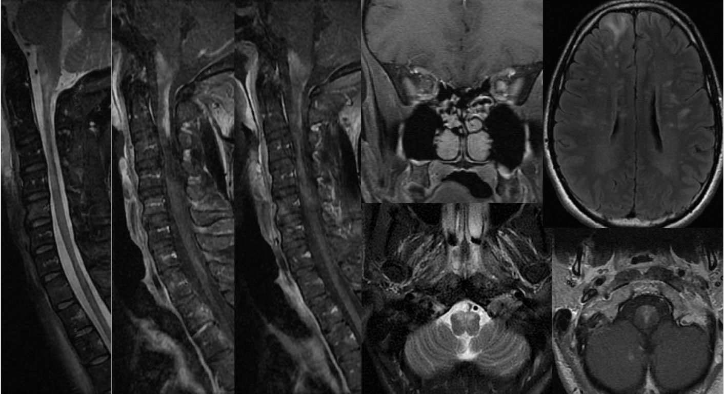
Six months later, addition of MRI cervical spine demonstrates extensive spinal cord signal abnormalities which are associated with patchy abnormal enhancement. The signal abnormalities are most concentrated in the upper to mid cervical cord, with the enhancement concentrated in the midline dorsal and ventral aspects of the upper cervical cord. There is also volume loss of the lower cervical cord. There is also a symmetric enhancement of the left optic nerve, with patchy signal abnormalities of the ventral medulla and progressive deep bilateral frontal white matter signal abnormalities. Additional signal abnormalities involve the bilateral anterior frontal lobes right greater than left, and there is additional abnormal enhancement within the medial right cerebellum.
Discussion:
The overall appearance of these white matter signal alterations suggest an underlying demyelinating process. However, the extensive patchy involvement of the cervical cord, as well as the enhancement pattern of the optic chiasm him and proximal optic nerves, with a generally midline distribution of the other signal abnormalities raise the possibility of neuromyelitis optica. The long segment diffuse involvement of the cervical cord and diffuse patchy enhancement is also unusual for multiple sclerosis and more typical for neuromyelitis optica.
BACK TO
MAIN PAGE