Metastatic Disease
Findings:
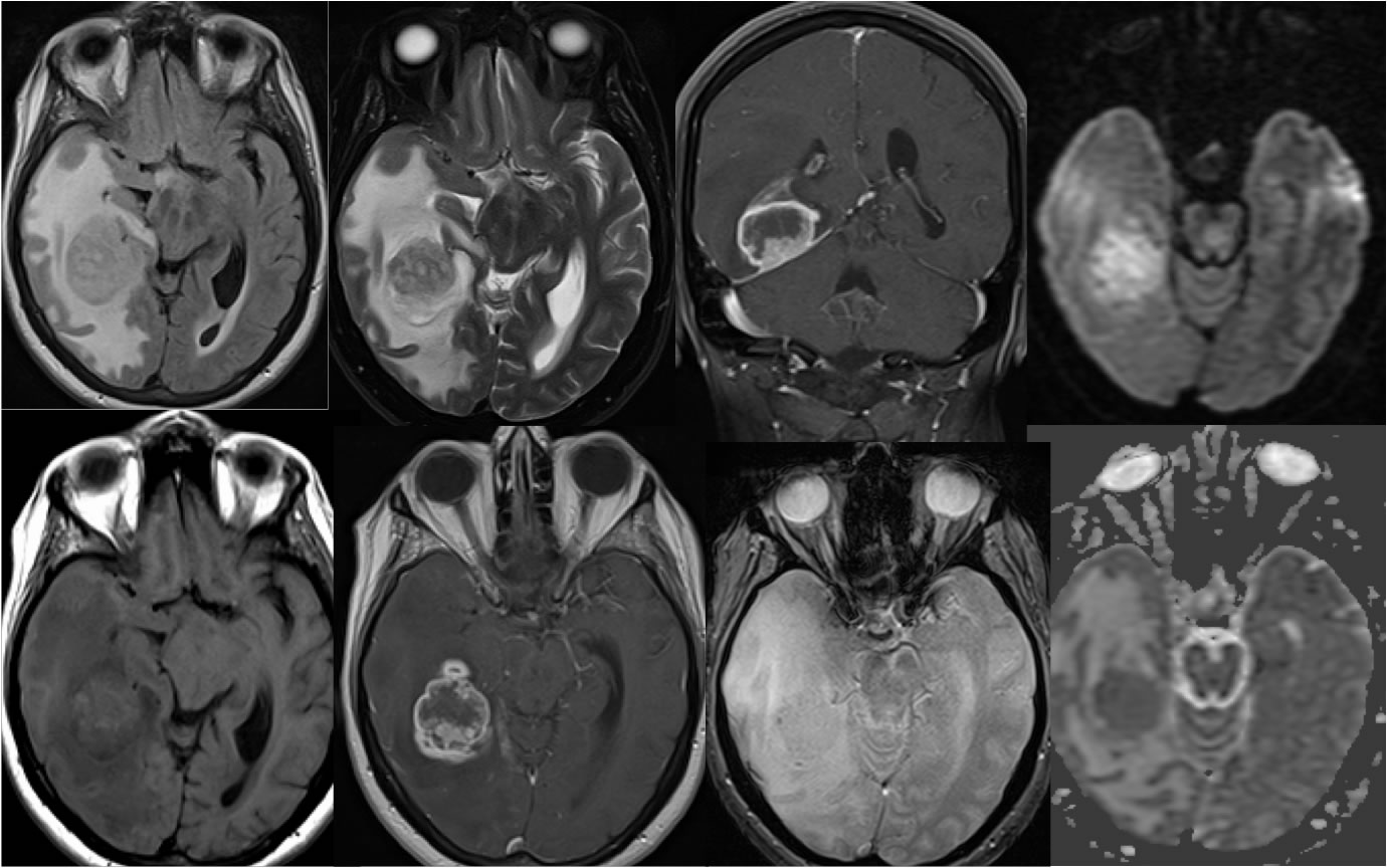
A heterogeneously enhancing mass is present in the right temporal lobe associated with extensive surrounding vasogenic edema. The mass demonstrate extensive nodular zones of peripheral enhancement, central necrosis, and no significant hemorrhagic change. The rim of the lesion is inconspicuous on T1 and T2 weighted imaging. The mass has a somewhat broad-based contact to the tentorium with associated mild linear dural thickening. The mass also demonstrates heterogeneous central restricted diffusion. The mass is inseparable from the right lateral ventricle.
Discussion:
While central restricted diffusion is invariably seen within cerebral abscesses, the diffusion signal in this case is less bright than typical for an abscess. The heterogeneous low signal of the mass on T2 weighted imaging is also a typical for abscess. The heterogeneous low signal is favored to represent complex necrotic fluid. The mass does have a broad contact to the dura, but overall appears extraaxial with no clear interface or displacement of cortex. The irregular nodularity and mass effect does not favor a demyelinating process. The top differential diagnostic considerations for this lesion include metastasis and glioblastoma.
BACK TO
MAIN PAGE