Multifocal Glioblastoma Multiforme
Findings:
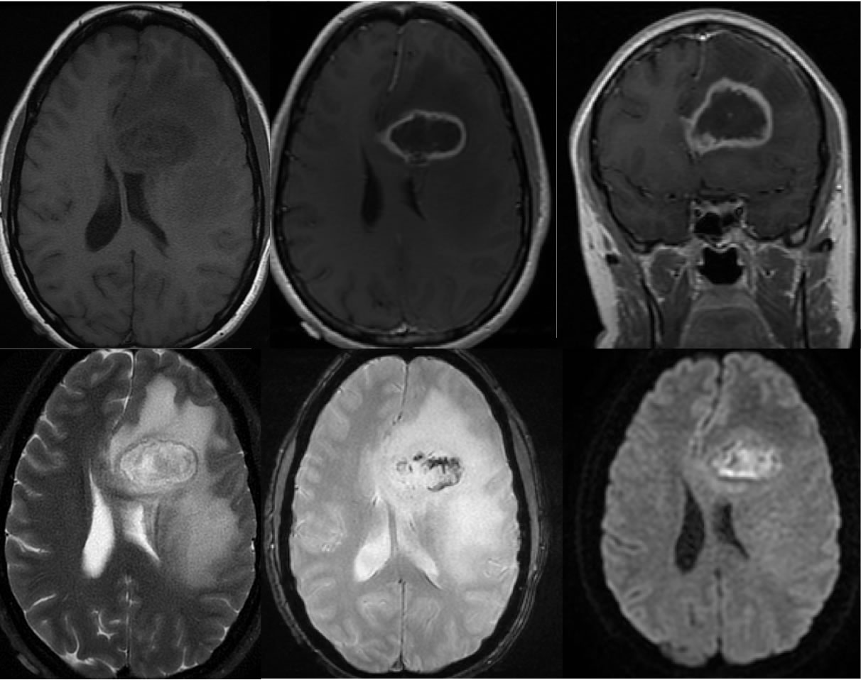
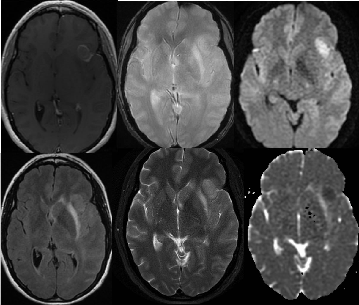
A large infiltrative peripherally enhancing mass with extensive surrounding signal alteration involves the left frontal lobe crossing the corpus callosum with associated mild to moderate mass effect, partial ventricular effacement, and mild right to left midline shift of the septum pellucidum. Gradient echo imaging demonstrates spotty hemorrhagic changes within the dominant legion. On diffusion weighted imaging, the necrotic hemorrhagic core of the lesion demonstrates relative heterogeneous increased signal. A second smaller poorly defined focus within the left inferior frontal lobe demonstrates minimal peripheral enhancement, prominent diffuse diffusion restriction, and no significant fluid component.
Discussion:
The central restricted diffusion of these lesions does mimic abscess, however the left inferior frontal lesion is not cystic/necrotic but is solid. The rounded fairly well defined nature of the left frontal lesion would be unusual for a focus of encephalitis. The central restricted diffusion of the dominant right frontal ring enhancing mass is also less bright than would be expected for an abscess, and may be partially accounted for by hemorrhagic changes visible on the GRE. An enhancing mass that crosses the corpus callosum has the primary differential diagnosis of glioblastoma.
BACK TO
MAIN PAGE