
Ruptured Basilar Terminus Aneurysm
Findings:
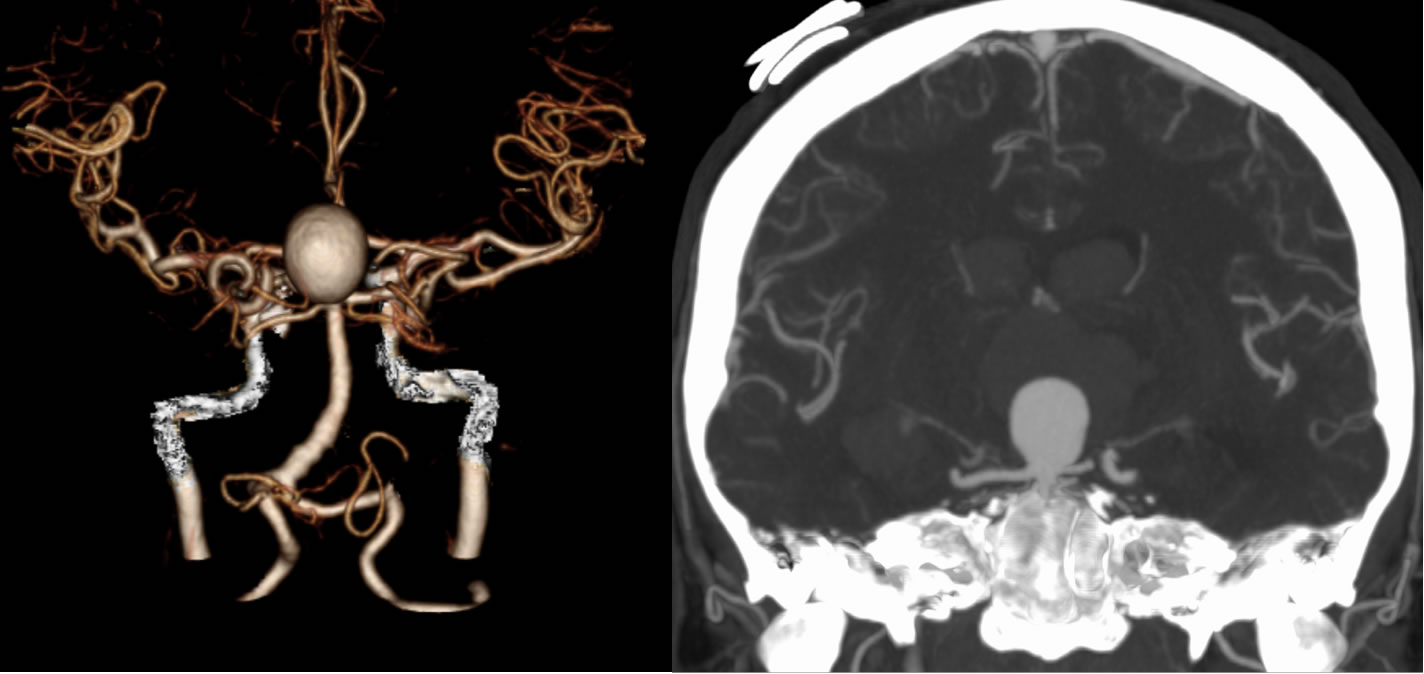
Axial noncontrast CT images demonstrate extensive intraventricular hemorrhage and scattered subarachnoid hemorrhage. A recently placed right frontal intraventricular catheter is incompletely included with pneumocephalus related to the procedure. The ventricular system is dilated. A rounded focus of relative decreased attenuation projects into the inferior third ventricle which correlates with the presence of a large basilar terminus aneurysm on the CTA.
Discussion:
The differential diagnosis for intracranial hemorrhage is very broad. The presence of subarachnoid hemorrhage should raise the possibility of a ruptured aneurysm. In this case, there is predominantly intraventricular hemorrhage which relates to rupture of the aneurysm directly into the ventricular system. Large aneurysms may be visible on noncontrast CT as rounded structures of relative lower attenuation floating in the surrounding hemorrhage. It is important to remember that the hyperdensity of intracranial hemorrhage relates to the coagulation, with flowing blood being of lower density than coagulated blood.
BACK TO
MAIN PAGE