Tumefactive Demyelination
Findings:
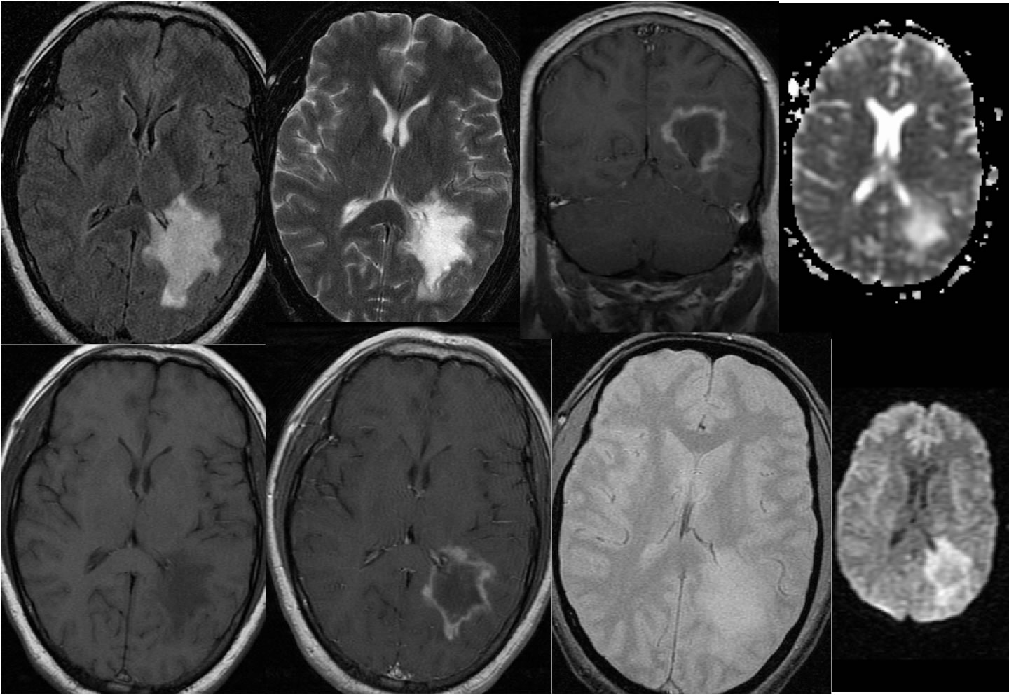
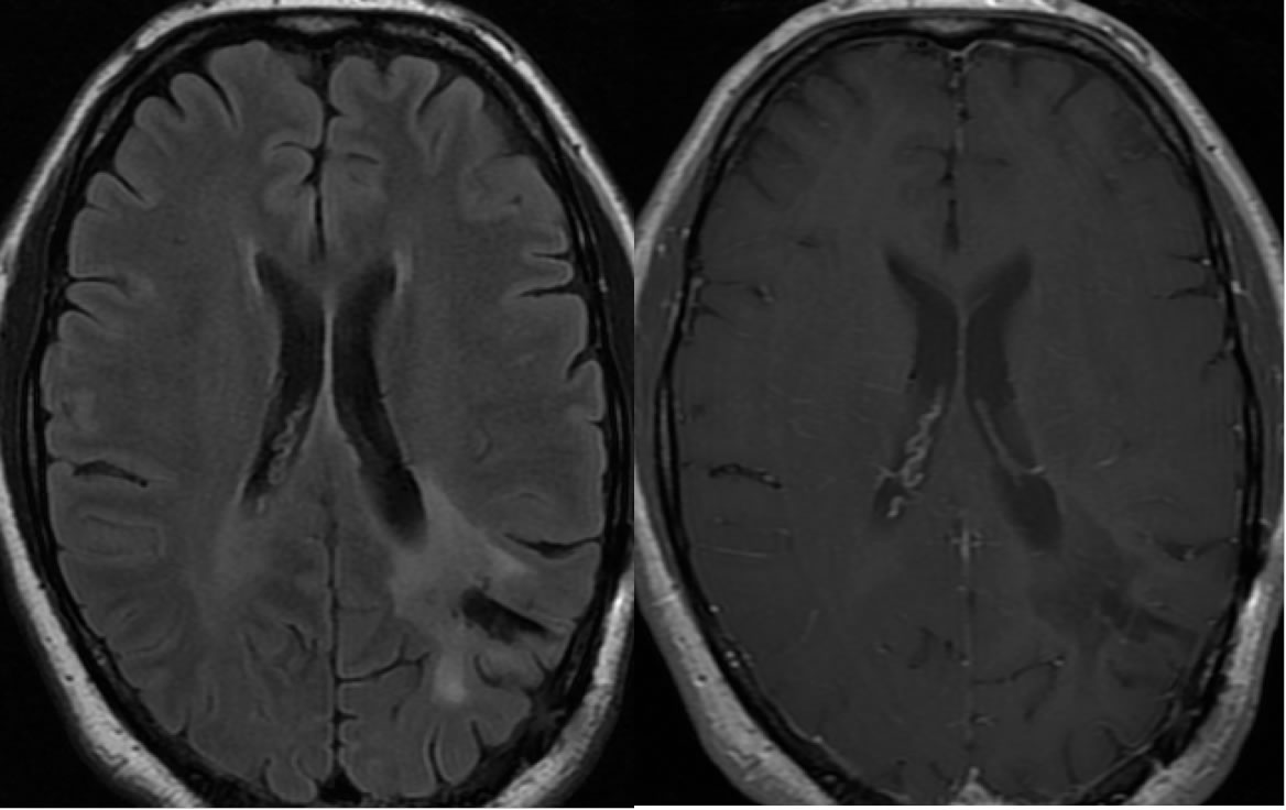
Multiple MR images demonstrate a large irregular zone of signal abnormality in the left parietooccipital lobe, associated with peripheral irregular enhancement. The peripheral irregular enhancement demonstrates relative restricted diffusion, with no central core of restricted diffusion. No hemorrhagic changes are seen. There is very little if any mass effect for the size of the lesion. On the follow up study, postoperative changes of craniotomy are now seen with a biopsy cavity within the previous lesion. The previous lesion has markedly improved on the follow-up examination.
Discussion:
The primary differential diagnostic considerations for this lesion include GBM and tumefactive demyelinating process. A solitary metastasis would be very unlikely in the absence of significant surrounding edema, and the signal characteristics are not compatible with abscess. One helpful feature to distinguish between GBM and tumefactive demyelination is the relative absence of signal abnormality peripheral to the advancing edge of enhancement, but not absolute since GBM may appear more well defined than the histology indicates. The irregular feathery morphology of the peripheral enhancement is more commonly seen with tumefactive demyelination than GBM, also not absolute. Peripheral diffusion restriction is similar to what would be seen with GBM. In many cases, biopsy is performed to differentiate between malignant brain tumor and large solitary tumefactive demyelinating process that simulates the appearance of neoplasm.
BACK TO
MAIN PAGE