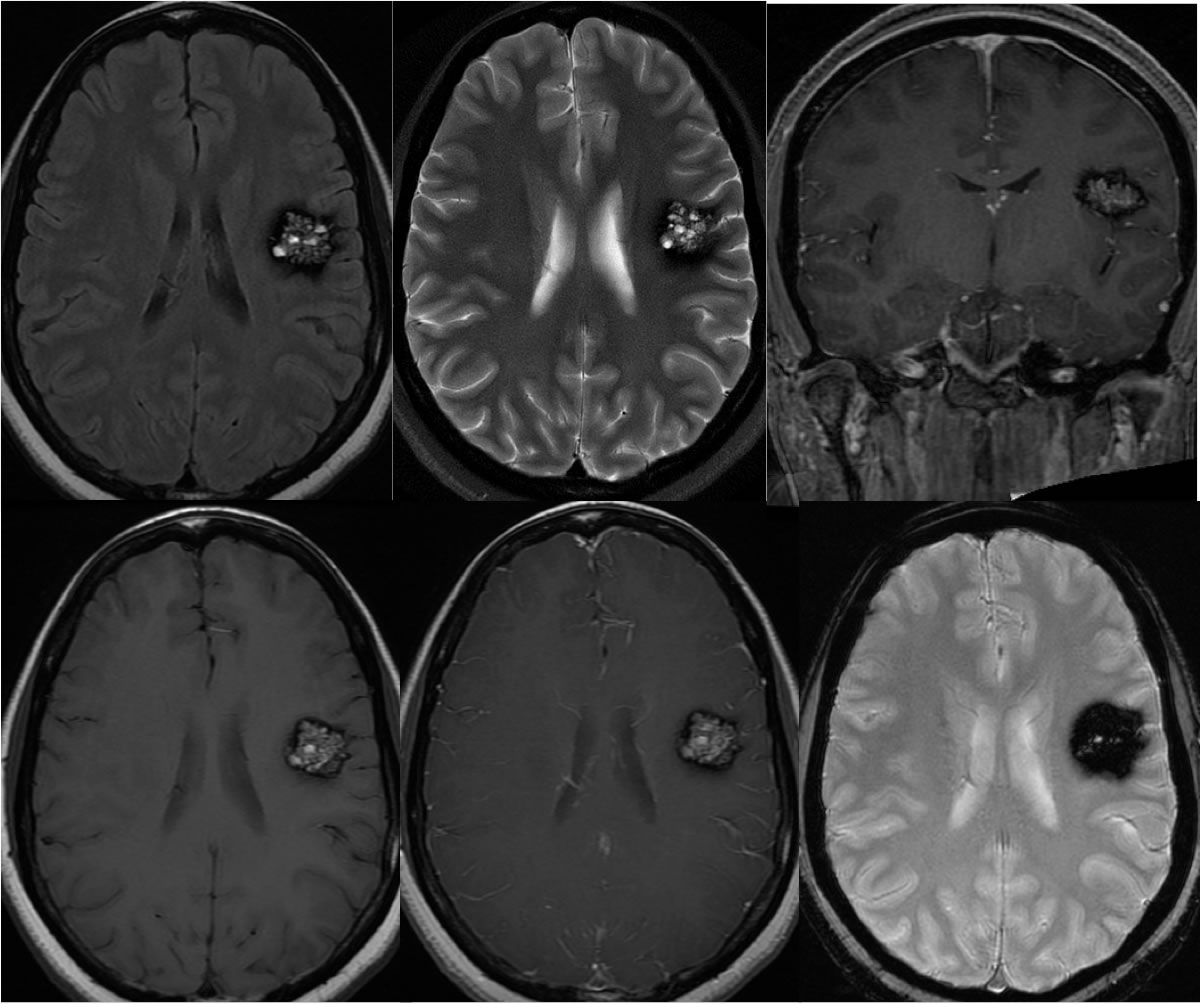
Cavernoma
Findings:
MR imaging at 3T demonstrates a rounded lesion within the subcortical white matter of the left frontal operculum, which is associated with a thick irregular peripheral hemosiderin rim and stippled zones of increased central T2 signal. The lesion demonstrates minimal enhancement after gadolinium administration. There is no associated mass effect or surrounding vasogenic edema.
Discussion:
This is a "classic" appearance for a cavernoma and other lesions are not considered likely to have this appearance. Occasionally, hemorrhagic metastatic or primary malignant neoplasms can share some features, but surrounding edema and mass effect would be expected.
BACK TO
MAIN PAGE