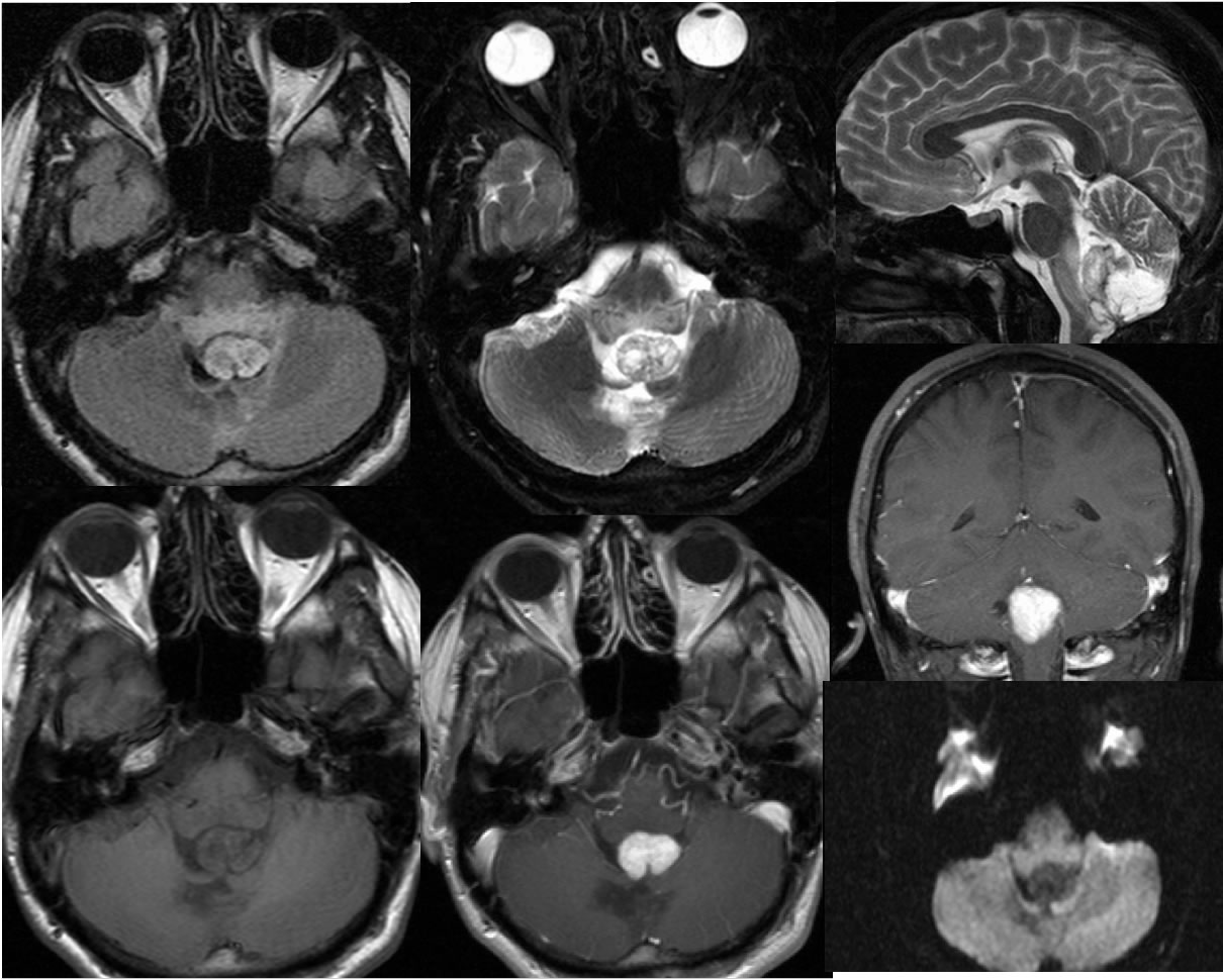
Recurrent Hemangioblastoma
Findings:
MR images demonstrate a strongly enhancing mass within the inferior aspect of the fourth ventricle. There is no diffusion restriction in this region. Surrounding vasogenic edema is present. Superimposed remote postoperative changes are seen within the cerebellar vermis. The mass demonstrates increased signal on FLAIR imaging and predominately hypointense signal on T1.
Differential Diagnosis:
Broad differential diagnosis, hemangioblastoma should be considered for an enhancing nodular cerebellar mass in an adult with no known primary cancer. Metastatic disease, medulloblastoma, and pilocytic astrocytoma can appear similar.
BACK TO
MAIN PAGE