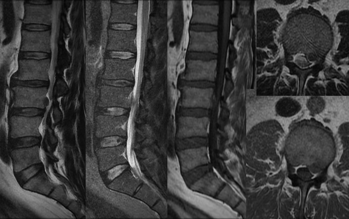
Sequestered Disc Extrusion L3-4
Findings:
Multiple MR images demonstrate a hypointense structure superior to the L3-L4 disc space on sagittal imaging. The lesion causes mild mass effect on the left thecal sac and demonstrates decreased T2 and intermediate T1 signal. Narrowing and dessication of the L2-3 disc is associated with osteophytes. Mild dessication and minimal subtle narrowing of the L3-4 disc space is present.
Discussion:
In the presence of degenerative disc disease, an extradural lesion associated with a degenerated disc that follows the signal of disc on all sequences represents a disc extrusion nearly always. In this case, the sequestered disc is located closer to L3-4 than L2-3, with the greatest degree of disc height loss and osteophytes at L2-3, therefore it is uncertain from which level the extrusion arises. The presence of an annular fissure could indicate the site of origin, but not definitely visible in this case. In the absence of degenerative disc disease, a T2 hypointense extradural mass could represent cellular neoplasm such as lymphoma or metastasis.
BACK TO
MAIN PAGE