CAPNON
Findings:
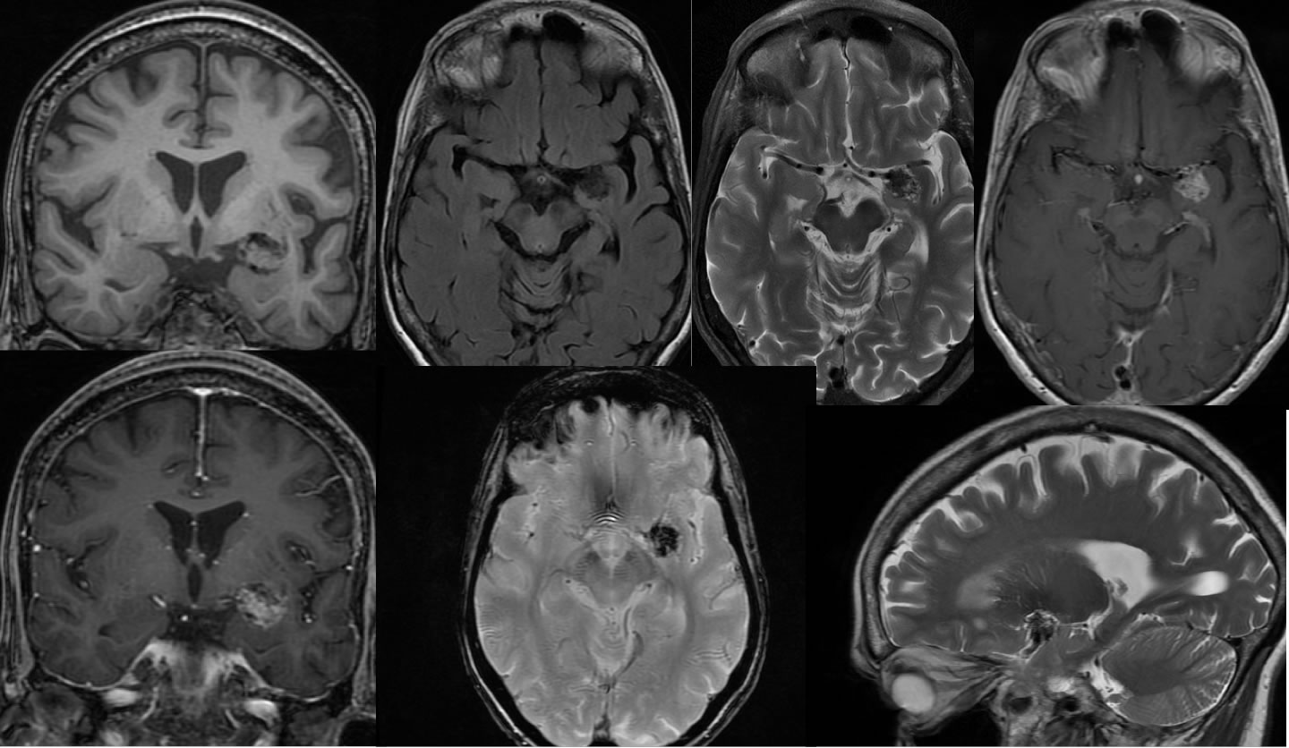
An irregular rounded focus of decreased T2 and decreased T1 signal involves the left medial temporal lobe, associated with abnormal enhancement, no mass effect, and no surrounding signal alteration. The mass has prominent hypointensity on the susceptibility weighted imaging suggestive of a calcified lesion.
Differential Diagnosis:
Features indicating a probable benign lesion include the lack of mass effect or surrounding edema. The enhancement pattern somewhat simulates meningioma, but this case is an intraaxial lesion. Due to proximity to vascular structures, a thrombosed vascular malformation might be considered. Some features simulate cavernoma, but the characteristic central T2 hyperintensity is not seen.
Discussion:
The differential diagnosis of an intracranial calcified lesion includes cavernous malformation, ganglioglioma, oligodendroglioma, meningioma, and choroid plexus papilloma. Calcifying pseudoneoplasm of the neuraxis (CAPNON) is rare with less than 30 cases reported, but the lesion does have fairly characteristic imaging features. On MR, these masses are isointense to hypointense on T1 weighted imaging, markedly hypointense on T2 imaging, and demonstrate variable enhancement with gradient echo blooming. The masses are most commonly extraaxial, but intraaxial masses are also seen and most common in the temporal lobe region. Patients typically present either asymptomatic or with seizures. Histologically, a chondromyxoid matrix is seen with extensive calcification and spindle cells that are EMA positive. The pathology may come back as a fibroosseous lesion. It is important to recognize the imaging features of these lesions to avoid more aggressive surgical or diagnostic interventions.
(Aiken, et al. AJNR June 2009 30:1256-1260).
BACK TO
MAIN PAGE