CADASIL
Findings:
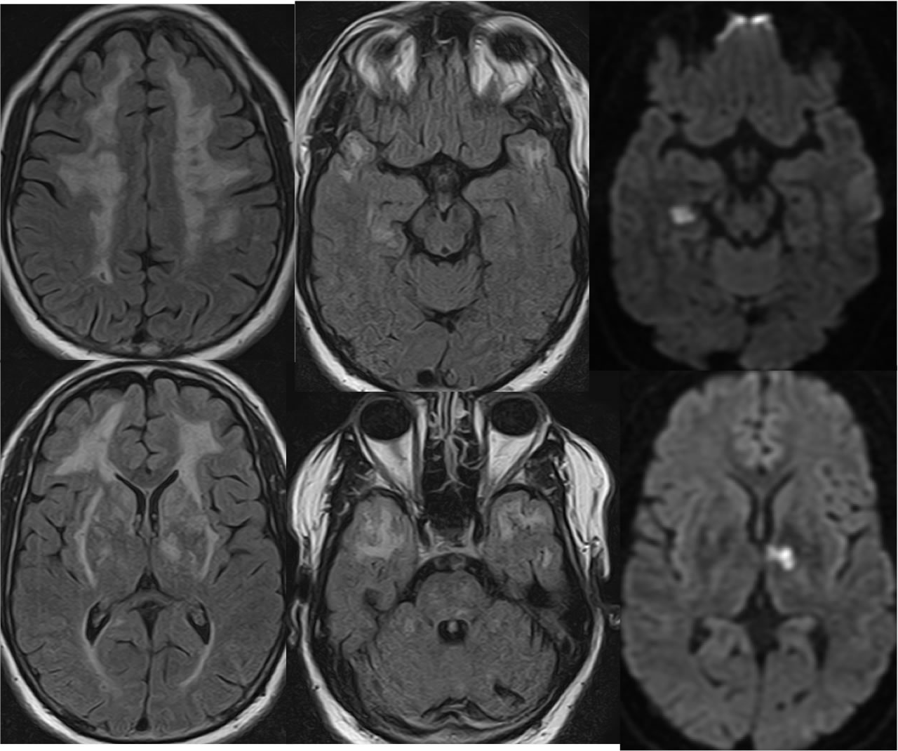
Axial FLAIR images show nearly symmetric signal abnormalities throughout the bilateral deep frontal, anterior temporal, and external capsule white matter tracts. Multiple remote lacunar infarcts involve the bilateral basal ganglia and left cerebellum. The diffusion weighted imaging demonstrates acute infarcts in the left thalamus and right medial temporal lobe.
Differential Diagnosis/Discussion:
The presence of extensive subcortical anterior temporal lobe white matter signal alterations raises the possibility of CADASIL over other causes of nonspecific white matter disease. While the age of the patient is not stated, a younger age group is suspected, with no significant cortical atrophy visible. While the process is somewhat periventricular, a characteristic ovoid morphology is not seen and there is no significant periatrial disease to suggest the possibility of demyelination.
BACK TO
MAIN PAGE