Sphenoid Sinus Mucocele and Incidental Meningioma
Findings:
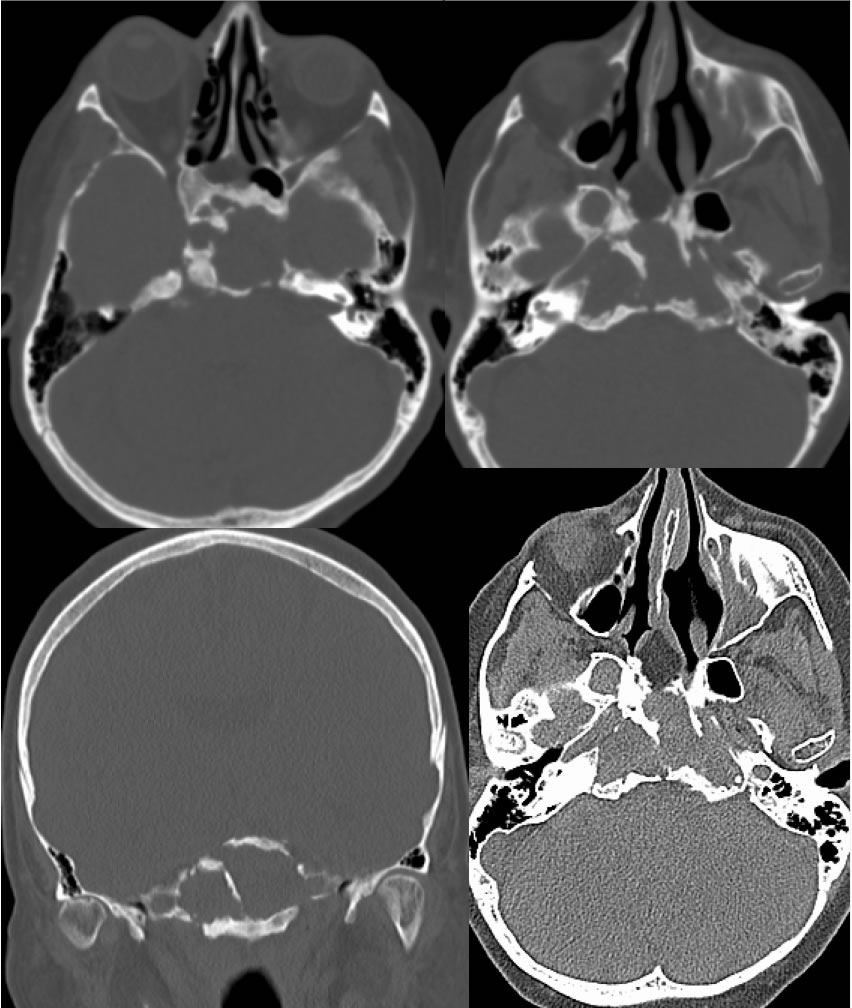
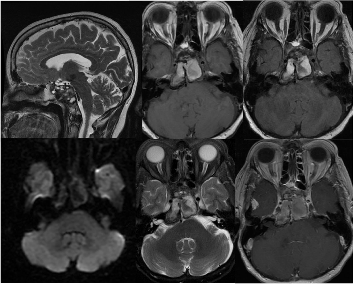
CT images show an irregular expansile process involving the sphenoid sinus with thickened sclerotic walls and multiple interruptions. The process is isodense to muscle and bone. Fat packing is seen along the nasal septum related to prior transsphenoidal surgery. MR images show mixed T2 and predominantly hyperintense T1 signal within this region, with no abnormal nodular enhancement. The sphenoid sinus process causes mild mass effect on the pons. An enhancing extraaxial mass extends along the right middle cranial fossa incompletely included.
Differential Diagnosis/Discussion:
An expansile lesion of the central skull base has a relatively broad differential diagnosis. With the fat packing along the posterior nasal septum, previous transsphenoidal procedure is confirmed. The lack of enhancement indicates a non-neoplastic process.
BACK TO
MAIN PAGE